 Movie
Movie Controller

[English] 日本語
 Yorodumi
- PDB-7t1q: Crystal Structure of the Succinyl-diaminopimelate Desuccinylase (... -
+ Open data

- Basic information
| Entry | Database: PDB / ID: 7t1q | ||||||
|---|---|---|---|---|---|---|---|
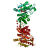
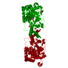
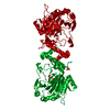
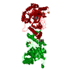
| Title | Crystal Structure of the Succinyl-diaminopimelate Desuccinylase (DapE) from Acinetobacter baumannii in complex with Succinic Acid | ||||||
 Components Components | Succinyl-diaminopimelate desuccinylase | ||||||
 Keywords Keywords | HYDROLASE / Structural Genomics / Center for Structural Genomics of Infectious Diseases / CSGID / DapE | ||||||
| Function / homology |  Function and homology information Function and homology informationsuccinyl-diaminopimelate desuccinylase / succinyl-diaminopimelate desuccinylase activity / acetylornithine deacetylase activity / L-arginine biosynthetic process / : / : / cobalt ion binding / zinc ion binding Similarity search - Function | ||||||
| Biological species |  Acinetobacter baumannii ATCC 17978 (bacteria) Acinetobacter baumannii ATCC 17978 (bacteria) | ||||||
| Method |  X-RAY DIFFRACTION / SYNCHROTRON / MOLECULAR REPLACEMENT / Resolution: 2.25 Å X-RAY DIFFRACTION / SYNCHROTRON / MOLECULAR REPLACEMENT / Resolution: 2.25 Å | ||||||
 Authors Authors | Minasov, G. / Shuvalova, L. / Brunzelle, J.S. / Dubrovska, I. / Pshenychnyi, S. / Satchell, K.J.F. / Center for Structural Genomics of Infectious Diseases (CSGID) | ||||||
| Funding support |  United States, 1items United States, 1items
| ||||||
 Citation Citation | Journal: Acs Omega / Year: 2024 Title: Biochemical and Structural Analysis of the Bacterial Enzyme Succinyl-Diaminopimelate Desuccinylase (DapE) from Acinetobacter baumannii. Authors: Kelley, E.H. / Minasov, G. / Konczak, K. / Shuvalova, L. / Brunzelle, J.S. / Shukla, S. / Beulke, M. / Thabthimthong, T. / Olsen, K.W. / Inniss, N.L. / Satchell, K.J.F. / Becker, D.P. | ||||||
| History |
|
- Structure visualization
| Structure viewer | Molecule: MolmilJmol/JSmol |
|---|
- Downloads & links
-Download
| PDBx/mmCIF format | 7t1q.cif.gz | 313.1 KB | Display | PDBx/mmCIF format |
|---|---|---|---|---|
| PDB format | pdb7t1q.ent.gz | 253.8 KB | Display | PDB format |
| PDBx/mmJSON format | 7t1q.json.gz | Tree view | PDBx/mmJSON format | |
| Others |  Other downloads Other downloads |
-Validation report
| Arichive directory | https://data.pdbj.org/pub/pdb/validation_reports/t1/7t1qftp://data.pdbj.org/pub/pdb/validation_reports/t1/7t1q | HTTPS FTP |
|---|
-Related structure data
| Related structure data |  8f8oC  5uejS S: Starting model for refinement C: citing same article ( |
|---|---|
| Similar structure data | |
| Other databases |


-Links
 PDBj
PDBj
- Assembly
| Deposited unit | 
| ||||||||
|---|---|---|---|---|---|---|---|---|---|
| 1 |
| ||||||||
| Unit cell |
|
-Components
-Protein , 1 types, 2 molecules AB
| #1: Protein | Mass: 41468.754 Da / Num. of mol.: 2 Source method: isolated from a genetically manipulated source Source: (gene. exp.) Acinetobacter baumannii ATCC 17978 (bacteria)Strain: ATCC 17978 / CIP 53.77 / LMG 1025 / NCDC KC755 / 5377 Gene: dapE, A1S_2810 / Plasmid: pMCSG92 / Production host: References: UniProt: A3M8H2, succinyl-diaminopimelate desuccinylase |
|---|
-Non-polymers , 5 types, 355 molecules 


| #2: Chemical |  Mass: 118.088 Da / Num. of mol.: 2 / Source method: obtained synthetically / Formula: C4H6O4 / Feature type: SUBJECT OF INVESTIGATION Mass: 118.088 Da / Num. of mol.: 2 / Source method: obtained synthetically / Formula: C4H6O4 / Feature type: SUBJECT OF INVESTIGATION#3: Chemical |  Mass: 59.044 Da / Num. of mol.: 2 / Source method: obtained synthetically / Formula: C2H3O2 Mass: 59.044 Da / Num. of mol.: 2 / Source method: obtained synthetically / Formula: C2H3O2#4: Chemical | ChemComp-PGE / |  Mass: 150.173 Da / Num. of mol.: 1 / Source method: obtained synthetically / Formula: C6H14O4 Mass: 150.173 Da / Num. of mol.: 1 / Source method: obtained synthetically / Formula: C6H14O4#5: Chemical | ChemComp-ZN /  Mass: 65.409 Da / Num. of mol.: 4 / Source method: obtained synthetically / Formula: Zn / Feature type: SUBJECT OF INVESTIGATION Mass: 65.409 Da / Num. of mol.: 4 / Source method: obtained synthetically / Formula: Zn / Feature type: SUBJECT OF INVESTIGATION#6: Water | ChemComp-HOH / | Mass: 18.015 Da / Num. of mol.: 346 / Source method: isolated from a natural source / Formula: H2O |
|---|
-Details
| Has ligand of interest | Y |
|---|---|
| Has protein modification | Y |
-Experimental details
-Experiment
| Experiment | Method: X-RAY DIFFRACTION / Number of used crystals: 1 |
|---|
- Sample preparation
| Crystal | Density Matthews: 2.94 Å3/Da / Density % sol: 58.6 % |
|---|---|
| Crystal grow | Temperature: 292 K / Method: vapor diffusion, sitting drop / pH: 4.5 Details: Protein: 10.6 mg/ml, 0.15M Sodium chloride, 0.01M Tris pH 8.3, 2mM N6-Me-L,L-SDAP; Screen: Classics II (D5), 0.1M Sodium acetate pH 4.5, 25% (w/v) PEG3350 |
-Data collection
| Diffraction | Mean temperature: 100 K / Serial crystal experiment: N |
|---|---|
| Diffraction source | Source: SYNCHROTRON / Site: APS / Beamline: 21-ID-D / Wavelength: 0.97848 Å |
| Detector | Type: DECTRIS EIGER X 9M / Detector: PIXEL / Date: Sep 17, 2021 / Details: mirrors |
| Radiation | Monochromator: Si / Protocol: SINGLE WAVELENGTH / Monochromatic (M) / Laue (L): M / Scattering type: x-ray |
| Radiation wavelength | Wavelength: 0.97848 Å / Relative weight: 1 |
| Reflection | Resolution: 2.25→30 Å / Num. obs: 50400 / % possible obs: 100 % / Observed criterion σ(I): -3 / Redundancy: 12.2 % / Biso Wilson estimate: 45.5 Å2 / CC1/2: 0.997 / CC star: 0.999 / Rmerge(I) obs: 0.145 / Rpim(I) all: 0.043 / Rrim(I) all: 0.151 / Rsym value: 0.145 / Χ2: 1.068 / Net I/σ(I): 20.2 |
| Reflection shell | Resolution: 2.25→2.29 Å / Redundancy: 12.5 % / Mean I/σ(I) obs: 1.7 / Num. unique obs: 2507 / CC1/2: 0.756 / CC star: 0.928 / Rpim(I) all: 0.607 / Χ2: 1.009 / % possible all: 100 |
- Processing
| Software |
| |||||||||||||||||||||||||||||||||||||||||||||||||||||||||||||||||||||||||||||||||||||||||||||||||||||||||||||||||||||||||||||||||||||||||||||||||||||||||||||||||||||||||||||||||||||||||||||||||||||||||||||||||||||||||||||||||
|---|---|---|---|---|---|---|---|---|---|---|---|---|---|---|---|---|---|---|---|---|---|---|---|---|---|---|---|---|---|---|---|---|---|---|---|---|---|---|---|---|---|---|---|---|---|---|---|---|---|---|---|---|---|---|---|---|---|---|---|---|---|---|---|---|---|---|---|---|---|---|---|---|---|---|---|---|---|---|---|---|---|---|---|---|---|---|---|---|---|---|---|---|---|---|---|---|---|---|---|---|---|---|---|---|---|---|---|---|---|---|---|---|---|---|---|---|---|---|---|---|---|---|---|---|---|---|---|---|---|---|---|---|---|---|---|---|---|---|---|---|---|---|---|---|---|---|---|---|---|---|---|---|---|---|---|---|---|---|---|---|---|---|---|---|---|---|---|---|---|---|---|---|---|---|---|---|---|---|---|---|---|---|---|---|---|---|---|---|---|---|---|---|---|---|---|---|---|---|---|---|---|---|---|---|---|---|---|---|---|---|---|---|---|---|---|---|---|---|---|---|---|---|---|---|---|---|
| Refinement | Method to determine structure: MOLECULAR REPLACEMENT Starting model: 5uej Resolution: 2.25→29.71 Å / Cor.coef. Fo:Fc: 0.965 / Cor.coef. Fo:Fc free: 0.944 / SU B: 14.067 / SU ML: 0.177 / Cross valid method: THROUGHOUT / σ(F): 0 / ESU R: 0.226 / ESU R Free: 0.194 / Stereochemistry target values: MAXIMUM LIKELIHOOD Details: HYDROGENS HAVE BEEN ADDED IN THE RIDING POSITIONS U VALUES : WITH TLS ADDED
| |||||||||||||||||||||||||||||||||||||||||||||||||||||||||||||||||||||||||||||||||||||||||||||||||||||||||||||||||||||||||||||||||||||||||||||||||||||||||||||||||||||||||||||||||||||||||||||||||||||||||||||||||||||||||||||||||
| Solvent computation | Ion probe radii: 0.8 Å / Shrinkage radii: 0.8 Å / VDW probe radii: 1.2 Å / Solvent model: MASK | |||||||||||||||||||||||||||||||||||||||||||||||||||||||||||||||||||||||||||||||||||||||||||||||||||||||||||||||||||||||||||||||||||||||||||||||||||||||||||||||||||||||||||||||||||||||||||||||||||||||||||||||||||||||||||||||||
| Displacement parameters | Biso max: 155.64 Å2 / Biso mean: 53.815 Å2 / Biso min: 22.46 Å2
| |||||||||||||||||||||||||||||||||||||||||||||||||||||||||||||||||||||||||||||||||||||||||||||||||||||||||||||||||||||||||||||||||||||||||||||||||||||||||||||||||||||||||||||||||||||||||||||||||||||||||||||||||||||||||||||||||
| Refinement step | Cycle: final / Resolution: 2.25→29.71 Å
| |||||||||||||||||||||||||||||||||||||||||||||||||||||||||||||||||||||||||||||||||||||||||||||||||||||||||||||||||||||||||||||||||||||||||||||||||||||||||||||||||||||||||||||||||||||||||||||||||||||||||||||||||||||||||||||||||
| Refine LS restraints |
| |||||||||||||||||||||||||||||||||||||||||||||||||||||||||||||||||||||||||||||||||||||||||||||||||||||||||||||||||||||||||||||||||||||||||||||||||||||||||||||||||||||||||||||||||||||||||||||||||||||||||||||||||||||||||||||||||
| LS refinement shell | Resolution: 2.25→2.308 Å / Rfactor Rfree error: 0 / Total num. of bins used: 20
| |||||||||||||||||||||||||||||||||||||||||||||||||||||||||||||||||||||||||||||||||||||||||||||||||||||||||||||||||||||||||||||||||||||||||||||||||||||||||||||||||||||||||||||||||||||||||||||||||||||||||||||||||||||||||||||||||
| Refinement TLS params. | Method: refined / Refine-ID: X-RAY DIFFRACTION
| |||||||||||||||||||||||||||||||||||||||||||||||||||||||||||||||||||||||||||||||||||||||||||||||||||||||||||||||||||||||||||||||||||||||||||||||||||||||||||||||||||||||||||||||||||||||||||||||||||||||||||||||||||||||||||||||||
| Refinement TLS group |
|
