 Movie
Movie Controller


+ Open data

- Basic information
| Entry | Database: PDB / ID: 7sv2 | ||||||
|---|---|---|---|---|---|---|---|
| Title | Human Cytochrome P450 (CYP) 3A5 ternary complex with azamulin | ||||||
 Components Components | Cytochrome P450 3A5 | ||||||
 Keywords Keywords | OXIDOREDUCTASE/OXIDOREDUCTASE INHIBITOR / Inhibitor / complex / binding properties / OXIDOREDUCTASE / OXIDOREDUCTASE-OXIDOREDUCTASE INHIBITOR complex | ||||||
| Function / homology |  Function and homology information Function and homology informationretinoic acid 4-hydroxylase activity / estrogen 16-alpha-hydroxylase activity / lipid hydroxylation / aflatoxin metabolic process / testosterone 6-beta-hydroxylase activity / alkaloid catabolic process / Aflatoxin activation and detoxification / estrogen 2-hydroxylase activity / oxidative demethylation / Xenobiotics ...retinoic acid 4-hydroxylase activity / estrogen 16-alpha-hydroxylase activity / lipid hydroxylation / aflatoxin metabolic process / testosterone 6-beta-hydroxylase activity / alkaloid catabolic process / Aflatoxin activation and detoxification / estrogen 2-hydroxylase activity / oxidative demethylation / Xenobiotics / retinoic acid metabolic process / estrogen metabolic process / retinol metabolic process / unspecific monooxygenase / steroid metabolic process / intracellular membrane-bounded organelle / xenobiotic catabolic process / xenobiotic metabolic process / monooxygenase activity / oxygen binding / oxidoreductase activity / iron ion binding / heme binding / endoplasmic reticulum membrane Similarity search - Function | ||||||
| Biological species |  Homo sapiens (human) Homo sapiens (human) | ||||||
| Method |  X-RAY DIFFRACTION / SYNCHROTRON / MOLECULAR REPLACEMENT / Resolution: 2.46 Å X-RAY DIFFRACTION / SYNCHROTRON / MOLECULAR REPLACEMENT / Resolution: 2.46 Å | ||||||
 Authors Authors | Hsu, M. / Johnson, E.F. | ||||||
| Funding support |  United States, 1items United States, 1items
| ||||||
 Citation Citation | Journal: J.Biol.Chem. / Year: 2022 Title: Structural characterization of the homotropic cooperative binding of azamulin to human cytochrome P450 3A5. Authors: Hsu, M.H. / Johnson, E.F. | ||||||
| History |
|
- Structure visualization
| Structure viewer | Molecule: MolmilJmol/JSmol |
|---|
- Downloads & links
-Download
| PDBx/mmCIF format | 7sv2.cif.gz | 886.6 KB | Display | PDBx/mmCIF format |
|---|---|---|---|---|
| PDB format | pdb7sv2.ent.gz | 618.3 KB | Display | PDB format |
| PDBx/mmJSON format | 7sv2.json.gz | Tree view | PDBx/mmJSON format | |
| Others |  Other downloads Other downloads |
-Validation report
| Arichive directory | https://data.pdbj.org/pub/pdb/validation_reports/sv/7sv2ftp://data.pdbj.org/pub/pdb/validation_reports/sv/7sv2 | HTTPS FTP |
|---|
-Related structure data
| Related structure data |  6mjmS S: Starting model for refinement |
|---|---|
| Similar structure data |
-Links
 PDBj
PDBj

- Assembly
| Deposited unit | 
| ||||||||||||
|---|---|---|---|---|---|---|---|---|---|---|---|---|---|
| 1 | 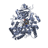
| ||||||||||||
| 2 | 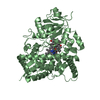
| ||||||||||||
| 3 | 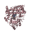
| ||||||||||||
| 4 | 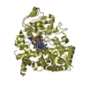
| ||||||||||||
| Unit cell |
|
-Components
| #1: Protein | Mass: 54862.586 Da / Num. of mol.: 4 / Fragment: UNP 24-497 Source method: isolated from a genetically manipulated source Source: (gene. exp.) Homo sapiens (human) / Gene: CYP3A5 / Plasmid: PCWORI / Production host:  #2: Chemical | ChemComp-HEM /   Mass: 616.487 Da / Num. of mol.: 4 / Source method: obtained synthetically / Formula: C34H32FeN4O4 / Feature type: SUBJECT OF INVESTIGATION Mass: 616.487 Da / Num. of mol.: 4 / Source method: obtained synthetically / Formula: C34H32FeN4O4 / Feature type: SUBJECT OF INVESTIGATION#3: Chemical | ChemComp-MWY / (   Mass: 478.648 Da / Num. of mol.: 8 / Source method: obtained synthetically / Formula: C24H38N4O4S Mass: 478.648 Da / Num. of mol.: 8 / Source method: obtained synthetically / Formula: C24H38N4O4S#4: Water | ChemComp-HOH / |  Mass: 18.015 Da / Num. of mol.: 128 / Source method: isolated from a natural source / Formula: H2O Mass: 18.015 Da / Num. of mol.: 128 / Source method: isolated from a natural source / Formula: H2OHas ligand of interest | Y | |
|---|
-Experimental details
-Experiment
| Experiment | Method: X-RAY DIFFRACTION / Number of used crystals: 1 |
|---|
- Sample preparation
| Crystal | Density Matthews: 2.21 Å3/Da / Density % sol: 44.26 % |
|---|---|
| Crystal grow | Temperature: 297 K / Method: vapor diffusion, sitting drop Details: Na Malonate, pH 6.5, 30% PEG 3350, n-decyl-beta-D-maltoside |
-Data collection
| Diffraction | Mean temperature: 100 K / Serial crystal experiment: N | ||||||||||||
|---|---|---|---|---|---|---|---|---|---|---|---|---|---|
| Diffraction source | Source: SYNCHROTRON / Site: SSRL / Beamline: BL12-1 / Wavelength: 0.97946 Å | ||||||||||||
| Detector | Type: DECTRIS EIGER X 16M / Detector: PIXEL / Date: Jul 8, 2020 | ||||||||||||
| Radiation | Protocol: SINGLE WAVELENGTH / Monochromatic (M) / Laue (L): M / Scattering type: x-ray | ||||||||||||
| Radiation wavelength | Wavelength: 0.97946 Å / Relative weight: 1 | ||||||||||||
| Reflection | Resolution: 2.3→39.56 Å / Num. obs: 85964 / % possible obs: 99.6 % / Redundancy: 12.1 % / Biso Wilson estimate: 50.98 Å2 / CC1/2: 0.996 / Net I/σ(I): 7.8 | ||||||||||||
| Reflection shell |
|
- Processing
| Software |
| |||||||||||||||||||||||||||||||||||||||||||||||||||||||||||||||||||||||||||||||||||||||||||||||||||||||||||||||||||||||||||||||||||||||||||||||||||||||||||||||||||||||||||||||
|---|---|---|---|---|---|---|---|---|---|---|---|---|---|---|---|---|---|---|---|---|---|---|---|---|---|---|---|---|---|---|---|---|---|---|---|---|---|---|---|---|---|---|---|---|---|---|---|---|---|---|---|---|---|---|---|---|---|---|---|---|---|---|---|---|---|---|---|---|---|---|---|---|---|---|---|---|---|---|---|---|---|---|---|---|---|---|---|---|---|---|---|---|---|---|---|---|---|---|---|---|---|---|---|---|---|---|---|---|---|---|---|---|---|---|---|---|---|---|---|---|---|---|---|---|---|---|---|---|---|---|---|---|---|---|---|---|---|---|---|---|---|---|---|---|---|---|---|---|---|---|---|---|---|---|---|---|---|---|---|---|---|---|---|---|---|---|---|---|---|---|---|---|---|---|---|---|
| Refinement | Method to determine structure: MOLECULAR REPLACEMENT Starting model: 6mjm Resolution: 2.46→39.44 Å / SU ML: 0.2865 / Cross valid method: FREE R-VALUE / σ(F): 1.35 / Phase error: 23.8963 Stereochemistry target values: GeoStd + Monomer Library + CDL v1.2
| |||||||||||||||||||||||||||||||||||||||||||||||||||||||||||||||||||||||||||||||||||||||||||||||||||||||||||||||||||||||||||||||||||||||||||||||||||||||||||||||||||||||||||||||
| Solvent computation | Shrinkage radii: 0.9 Å / VDW probe radii: 1.11 Å / Solvent model: FLAT BULK SOLVENT MODEL | |||||||||||||||||||||||||||||||||||||||||||||||||||||||||||||||||||||||||||||||||||||||||||||||||||||||||||||||||||||||||||||||||||||||||||||||||||||||||||||||||||||||||||||||
| Displacement parameters | Biso mean: 63.54 Å2 | |||||||||||||||||||||||||||||||||||||||||||||||||||||||||||||||||||||||||||||||||||||||||||||||||||||||||||||||||||||||||||||||||||||||||||||||||||||||||||||||||||||||||||||||
| Refinement step | Cycle: LAST / Resolution: 2.46→39.44 Å
| |||||||||||||||||||||||||||||||||||||||||||||||||||||||||||||||||||||||||||||||||||||||||||||||||||||||||||||||||||||||||||||||||||||||||||||||||||||||||||||||||||||||||||||||
| Refine LS restraints |
| |||||||||||||||||||||||||||||||||||||||||||||||||||||||||||||||||||||||||||||||||||||||||||||||||||||||||||||||||||||||||||||||||||||||||||||||||||||||||||||||||||||||||||||||
| LS refinement shell |
| |||||||||||||||||||||||||||||||||||||||||||||||||||||||||||||||||||||||||||||||||||||||||||||||||||||||||||||||||||||||||||||||||||||||||||||||||||||||||||||||||||||||||||||||
| Refinement TLS params. | Method: refined / Refine-ID: X-RAY DIFFRACTION
| |||||||||||||||||||||||||||||||||||||||||||||||||||||||||||||||||||||||||||||||||||||||||||||||||||||||||||||||||||||||||||||||||||||||||||||||||||||||||||||||||||||||||||||||
| Refinement TLS group | Refine-ID: X-RAY DIFFRACTION
|
