 ムービー
ムービー コントローラー


+ データを開く

- 基本情報
基本情報
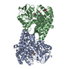
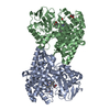
| 登録情報 | データベース: PDB / ID: 7stv | ||||||
|---|---|---|---|---|---|---|---|
| タイトル | Crystal structure of sulfatase from Pedobacter yulinensis | ||||||
 要素 要素 | N-acetylgalactosamine-6-sulfatase | ||||||
 キーワード キーワード | HYDROLASE | ||||||
| 機能・相同性 |  機能・相同性情報 機能・相同性情報 | ||||||
| 生物種 |  Pedobacter yulinensis (バクテリア) Pedobacter yulinensis (バクテリア) | ||||||
| 手法 |  X線回折 / シンクロトロン / 分子置換 / 解像度: 2.35 Å X線回折 / シンクロトロン / 分子置換 / 解像度: 2.35 Å | ||||||
 データ登録者 データ登録者 | O'Malley, A. / Schlachter, C.R. / Grimes, L.L. / Tomashek, J.J. / Lee, A.L. / Chruszcz, M. | ||||||
| 資金援助 | 1件
| ||||||
 引用 引用 | ジャーナル: Molecules / 年: 2021 タイトル: Purification, Characterization, and Structural Studies of a Sulfatase from Pedobacter yulinensis . 著者: Schlachter, C.R. / O'Malley, A. / Grimes, L.L. / Tomashek, J.J. / Chruszcz, M. / Lee, L.A. | ||||||
| 履歴 |
|
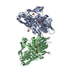
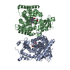
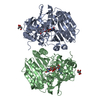
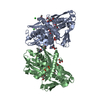
- 構造の表示
構造の表示
| 構造ビューア | 分子: MolmilJmol/JSmol |
|---|
- ダウンロードとリンク
-ダウンロード
| PDBx/mmCIF形式 | 7stv.cif.gz | 179 KB | 表示 | PDBx/mmCIF形式 |
|---|---|---|---|---|
| PDB形式 | pdb7stv.ent.gz | 135.4 KB | 表示 | PDB形式 |
| PDBx/mmJSON形式 | 7stv.json.gz | ツリー表示 | PDBx/mmJSON形式 | |
| その他 |  その他のダウンロード その他のダウンロード |
-検証レポート
| 文書・要旨 | 7stv_validation.pdf.gz | 852.2 KB | 表示 | wwPDB検証レポート |
|---|---|---|---|---|
| 文書・詳細版 | 7stv_full_validation.pdf.gz | 857.8 KB | 表示 | |
| XML形式データ | 7stv_validation.xml.gz | 18.8 KB | 表示 | |
| CIF形式データ | 7stv_validation.cif.gz | 26.2 KB | 表示 | |
| アーカイブディレクトリ | https://data.pdbj.org/pub/pdb/validation_reports/st/7stvftp://data.pdbj.org/pub/pdb/validation_reports/st/7stv | HTTPS FTP |
-関連構造データ


-リンク
 PDBj
PDBj
- 集合体
集合体
| 登録構造単位 | 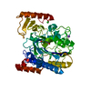
| |||||||||
|---|---|---|---|---|---|---|---|---|---|---|
| 1 | 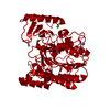
| |||||||||
| 単位格子 |
| |||||||||
| Components on special symmetry positions |
|
-要素
-タンパク質 , 1種, 1分子 A
| #1: タンパク質 | 分子量: 51392.445 Da / 分子数: 1 / 由来タイプ: 組換発現 由来: (組換発現) Pedobacter yulinensis (バクテリア)遺伝子: C7T94_09545 / 発現宿主: |
|---|
-非ポリマー , 5種, 122分子 


| #2: 化合物 | ChemComp-CIT /  分子量: 192.124 Da / 分子数: 1 / 由来タイプ: 合成 / 式: C6H8O7 分子量: 192.124 Da / 分子数: 1 / 由来タイプ: 合成 / 式: C6H8O7 | ||||||
|---|---|---|---|---|---|---|---|
| #3: 化合物 |  分子量: 22.990 Da / 分子数: 2 / 由来タイプ: 合成 / 式: Na 分子量: 22.990 Da / 分子数: 2 / 由来タイプ: 合成 / 式: Na#4: 化合物 |  分子量: 35.453 Da / 分子数: 2 / 由来タイプ: 合成 / 式: Cl 分子量: 35.453 Da / 分子数: 2 / 由来タイプ: 合成 / 式: Cl#5: 化合物 | ChemComp-CA / |  分子量: 40.078 Da / 分子数: 1 / 由来タイプ: 合成 / 式: Ca / タイプ: SUBJECT OF INVESTIGATION 分子量: 40.078 Da / 分子数: 1 / 由来タイプ: 合成 / 式: Ca / タイプ: SUBJECT OF INVESTIGATION#6: 水 | ChemComp-HOH / | 分子量: 18.015 Da / 分子数: 116 / 由来タイプ: 天然 / 式: H2O |
-詳細
| 研究の焦点であるリガンドがあるか | Y |
|---|---|
| Has protein modification | Y |
-実験情報
-実験
| 実験 | 手法: X線回折 / 使用した結晶の数: 1 |
|---|
- 試料調製
試料調製
| 結晶 | マシュー密度: 2.22 Å3/Da / 溶媒含有率: 44.5 % |
|---|---|
| 結晶化 | 温度: 295 K / 手法: 蒸気拡散法, シッティングドロップ法 / pH: 5.5 詳細: 0.1 M sodium citrate pH 5.5, 20% w/v polyethylene glycol 3000 |
-データ収集
| 回折 | 平均測定温度: 295 K / Serial crystal experiment: N |
|---|---|
| 放射光源 | 由来: シンクロトロン / サイト: APS  / ビームライン: 22-ID / 波長: 1 Å / ビームライン: 22-ID / 波長: 1 Å |
| 検出器 | タイプ: DECTRIS EIGER X 16M / 検出器: PIXEL / 日付: 2021年6月29日 |
| 放射 | プロトコル: SINGLE WAVELENGTH / 単色(M)・ラウエ(L): M / 散乱光タイプ: x-ray |
| 放射波長 | 波長: 1 Å / 相対比: 1 |
| 反射 | 解像度: 2.34→40 Å / Num. obs: 19275 / % possible obs: 98.8 % / 冗長度: 9.2 % / CC1/2: 0.998 / CC star: 0.999 / Rmerge(I) obs: 0.101 / Rpim(I) all: 0.048 / Rrim(I) all: 0.148 / Rsym value: 0.101 / Net I/σ(I): 18.5 |
| 反射 シェル | 解像度: 2.34→2.38 Å / 冗長度: 6.7 % / Rmerge(I) obs: 0.489 / Mean I/σ(I) obs: 2 / Num. unique obs: 936 / CC1/2: 0.948 / CC star: 0.986 / Rpim(I) all: 0.175 / Rrim(I) all: 0.525 / Rsym value: 0.489 / % possible all: 98.7 |
- 解析
解析
| ソフトウェア |
| |||||||||||||||||||||||||||||||||||||||||||||||||||||||||||||||||||||||||||||||||||||||||||||||||||||||||||||||||||||||||||||||||||||||||||||||||||||||||||||||||||||
|---|---|---|---|---|---|---|---|---|---|---|---|---|---|---|---|---|---|---|---|---|---|---|---|---|---|---|---|---|---|---|---|---|---|---|---|---|---|---|---|---|---|---|---|---|---|---|---|---|---|---|---|---|---|---|---|---|---|---|---|---|---|---|---|---|---|---|---|---|---|---|---|---|---|---|---|---|---|---|---|---|---|---|---|---|---|---|---|---|---|---|---|---|---|---|---|---|---|---|---|---|---|---|---|---|---|---|---|---|---|---|---|---|---|---|---|---|---|---|---|---|---|---|---|---|---|---|---|---|---|---|---|---|---|---|---|---|---|---|---|---|---|---|---|---|---|---|---|---|---|---|---|---|---|---|---|---|---|---|---|---|---|---|---|---|---|---|
| 精密化 | 構造決定の手法: 分子置換 開始モデル: 7STT 解像度: 2.35→34.232 Å / Cor.coef. Fo:Fc: 0.967 / Cor.coef. Fo:Fc free: 0.937 / SU B: 19.712 / SU ML: 0.209 / 交差検証法: FREE R-VALUE / ESU R: 0.393 / ESU R Free: 0.244 / 詳細: Hydrogens have been added in their riding positions
| |||||||||||||||||||||||||||||||||||||||||||||||||||||||||||||||||||||||||||||||||||||||||||||||||||||||||||||||||||||||||||||||||||||||||||||||||||||||||||||||||||||
| 溶媒の処理 | イオンプローブ半径: 0.8 Å / 減衰半径: 0.8 Å / VDWプローブ半径: 1.2 Å / 溶媒モデル: MASK BULK SOLVENT | |||||||||||||||||||||||||||||||||||||||||||||||||||||||||||||||||||||||||||||||||||||||||||||||||||||||||||||||||||||||||||||||||||||||||||||||||||||||||||||||||||||
| 原子変位パラメータ | Biso mean: 56.402 Å2
| |||||||||||||||||||||||||||||||||||||||||||||||||||||||||||||||||||||||||||||||||||||||||||||||||||||||||||||||||||||||||||||||||||||||||||||||||||||||||||||||||||||
| 精密化ステップ | サイクル: LAST / 解像度: 2.35→34.232 Å
| |||||||||||||||||||||||||||||||||||||||||||||||||||||||||||||||||||||||||||||||||||||||||||||||||||||||||||||||||||||||||||||||||||||||||||||||||||||||||||||||||||||
| 拘束条件 |
| |||||||||||||||||||||||||||||||||||||||||||||||||||||||||||||||||||||||||||||||||||||||||||||||||||||||||||||||||||||||||||||||||||||||||||||||||||||||||||||||||||||
| LS精密化 シェル |
| |||||||||||||||||||||||||||||||||||||||||||||||||||||||||||||||||||||||||||||||||||||||||||||||||||||||||||||||||||||||||||||||||||||||||||||||||||||||||||||||||||||
| 精密化 TLS | 手法: refined / Origin x: -33.34 Å / Origin y: 6.47 Å / Origin z: -16.9122 Å
| |||||||||||||||||||||||||||||||||||||||||||||||||||||||||||||||||||||||||||||||||||||||||||||||||||||||||||||||||||||||||||||||||||||||||||||||||||||||||||||||||||||
| 精密化 TLSグループ | Selection: ALL |
