 Movie
Movie Controller


+ Open data

- Basic information
| Entry | Database: PDB / ID: 7stu | ||||||
|---|---|---|---|---|---|---|---|
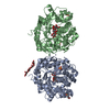
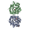
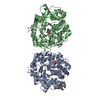
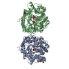
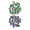
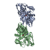
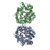
| Title | Crystal structure of sulfatase from Pedobacter yulinensis | ||||||
 Components Components | N-acetylgalactosamine-6-sulfatase | ||||||
 Keywords Keywords | HYDROLASE | ||||||
| Function / homology |  Function and homology information Function and homology information | ||||||
| Biological species |  Pedobacter yulinensis (bacteria) Pedobacter yulinensis (bacteria) | ||||||
| Method |  X-RAY DIFFRACTION / SYNCHROTRON / MOLECULAR REPLACEMENT / Resolution: 2.23 Å X-RAY DIFFRACTION / SYNCHROTRON / MOLECULAR REPLACEMENT / Resolution: 2.23 Å | ||||||
 Authors Authors | O'Malley, A. / Schlachter, C.R. / Grimes, L.L. / Tomashek, J.J. / Lee, A.L. / Chruszcz, M. | ||||||
| Funding support | 1items
| ||||||
 Citation Citation | Journal: Molecules / Year: 2021 Title: Purification, Characterization, and Structural Studies of a Sulfatase from Pedobacter yulinensis . Authors: Schlachter, C.R. / O'Malley, A. / Grimes, L.L. / Tomashek, J.J. / Chruszcz, M. / Lee, L.A. | ||||||
| History |
|
- Structure visualization
| Structure viewer | Molecule: MolmilJmol/JSmol |
|---|
- Downloads & links
-Download
| PDBx/mmCIF format | 7stu.cif.gz | 180.3 KB | Display | PDBx/mmCIF format |
|---|---|---|---|---|
| PDB format | pdb7stu.ent.gz | 136.8 KB | Display | PDB format |
| PDBx/mmJSON format | 7stu.json.gz | Tree view | PDBx/mmJSON format | |
| Others |  Other downloads Other downloads |
-Validation report
| Summary document | 7stu_validation.pdf.gz | 837.6 KB | Display | wwPDB validaton report |
|---|---|---|---|---|
| Full document | 7stu_full_validation.pdf.gz | 839.3 KB | Display | |
| Data in XML | 7stu_validation.xml.gz | 20.6 KB | Display | |
| Data in CIF | 7stu_validation.cif.gz | 30.8 KB | Display | |
| Arichive directory | https://data.pdbj.org/pub/pdb/validation_reports/st/7stuftp://data.pdbj.org/pub/pdb/validation_reports/st/7stu | HTTPS FTP |
-Related structure data
| Related structure data |  7sttSC  7stvC S: Starting model for refinement C: citing same article ( |
|---|---|
| Similar structure data |


-Links
 PDBj
PDBj


- Assembly
| Deposited unit | 
| ||||||||
|---|---|---|---|---|---|---|---|---|---|
| 1 | 
| ||||||||
| Unit cell |
| ||||||||
| Components on special symmetry positions |
|
-Components
| #1: Protein | Mass: 51392.445 Da / Num. of mol.: 1 Source method: isolated from a genetically manipulated source Source: (gene. exp.) Pedobacter yulinensis (bacteria) / Gene: C7T94_09545 / Production host: | ||||||||||
|---|---|---|---|---|---|---|---|---|---|---|---|
| #2: Chemical | ChemComp-BR /   Mass: 79.904 Da / Num. of mol.: 5 / Source method: obtained synthetically / Formula: Br Mass: 79.904 Da / Num. of mol.: 5 / Source method: obtained synthetically / Formula: Br#3: Chemical |   Mass: 22.990 Da / Num. of mol.: 3 / Source method: obtained synthetically / Formula: Na Mass: 22.990 Da / Num. of mol.: 3 / Source method: obtained synthetically / Formula: Na#4: Chemical | ChemComp-CA / |   Mass: 40.078 Da / Num. of mol.: 1 / Source method: obtained synthetically / Formula: Ca / Feature type: SUBJECT OF INVESTIGATION Mass: 40.078 Da / Num. of mol.: 1 / Source method: obtained synthetically / Formula: Ca / Feature type: SUBJECT OF INVESTIGATION#5: Water | ChemComp-HOH / |  Mass: 18.015 Da / Num. of mol.: 316 / Source method: isolated from a natural source / Formula: H2O Mass: 18.015 Da / Num. of mol.: 316 / Source method: isolated from a natural source / Formula: H2OHas ligand of interest | Y | Has protein modification | Y | |
-Experimental details
-Experiment
| Experiment | Method: X-RAY DIFFRACTION / Number of used crystals: 1 |
|---|
- Sample preparation
| Crystal | Density Matthews: 2.26 Å3/Da / Density % sol: 45.47 % |
|---|---|
| Crystal grow | Temperature: 295 K / Method: vapor diffusion, sitting drop Details: 0.15 M potassium bromide, 30% w/v polyethylene glycol monomethyl ether 2000 |
-Data collection
| Diffraction | Mean temperature: 295 K / Serial crystal experiment: N |
|---|---|
| Diffraction source | Source: SYNCHROTRON / Site: APS  / Beamline: 22-ID / Wavelength: 1 Å / Beamline: 22-ID / Wavelength: 1 Å |
| Detector | Type: DECTRIS EIGER X 16M / Detector: PIXEL / Date: Jun 29, 2021 |
| Radiation | Protocol: SINGLE WAVELENGTH / Monochromatic (M) / Laue (L): M / Scattering type: x-ray |
| Radiation wavelength | Wavelength: 1 Å / Relative weight: 1 |
| Reflection | Resolution: 2.23→40 Å / Num. obs: 23149 / % possible obs: 99.2 % / Redundancy: 9.1 % / CC1/2: 0.997 / CC star: 0.999 / Rmerge(I) obs: 0.128 / Rpim(I) all: 0.057 / Rrim(I) all: 0.172 / Rsym value: 0.128 / Net I/σ(I): 15.14 |
| Reflection shell | Resolution: 2.23→2.27 Å / Redundancy: 6.2 % / Rmerge(I) obs: 0.907 / Mean I/σ(I) obs: 2 / Num. unique obs: 1144 / CC1/2: 0.738 / CC star: 0.922 / Rpim(I) all: 0.339 / Rrim(I) all: 0.858 / Rsym value: 0.907 / % possible all: 99.6 |
- Processing
| Software |
| |||||||||||||||||||||||||||||||||||||||||||||||||||||||||||||||||||||||||||||||||||||||||||||||||||||||||||||||||||||||||||||||||||||||||||||||||||||||||||||||||||||
|---|---|---|---|---|---|---|---|---|---|---|---|---|---|---|---|---|---|---|---|---|---|---|---|---|---|---|---|---|---|---|---|---|---|---|---|---|---|---|---|---|---|---|---|---|---|---|---|---|---|---|---|---|---|---|---|---|---|---|---|---|---|---|---|---|---|---|---|---|---|---|---|---|---|---|---|---|---|---|---|---|---|---|---|---|---|---|---|---|---|---|---|---|---|---|---|---|---|---|---|---|---|---|---|---|---|---|---|---|---|---|---|---|---|---|---|---|---|---|---|---|---|---|---|---|---|---|---|---|---|---|---|---|---|---|---|---|---|---|---|---|---|---|---|---|---|---|---|---|---|---|---|---|---|---|---|---|---|---|---|---|---|---|---|---|---|---|
| Refinement | Method to determine structure: MOLECULAR REPLACEMENT Starting model: 7STT Resolution: 2.23→39.217 Å / Cor.coef. Fo:Fc: 0.957 / Cor.coef. Fo:Fc free: 0.923 / SU B: 14.177 / SU ML: 0.164 / Cross valid method: FREE R-VALUE / ESU R: 0.302 / ESU R Free: 0.215 Details: Hydrogens have been added in their riding positions
| |||||||||||||||||||||||||||||||||||||||||||||||||||||||||||||||||||||||||||||||||||||||||||||||||||||||||||||||||||||||||||||||||||||||||||||||||||||||||||||||||||||
| Solvent computation | Ion probe radii: 0.8 Å / Shrinkage radii: 0.8 Å / VDW probe radii: 1.2 Å / Solvent model: BABINET MODEL PLUS MASK | |||||||||||||||||||||||||||||||||||||||||||||||||||||||||||||||||||||||||||||||||||||||||||||||||||||||||||||||||||||||||||||||||||||||||||||||||||||||||||||||||||||
| Displacement parameters | Biso mean: 33.47 Å2
| |||||||||||||||||||||||||||||||||||||||||||||||||||||||||||||||||||||||||||||||||||||||||||||||||||||||||||||||||||||||||||||||||||||||||||||||||||||||||||||||||||||
| Refinement step | Cycle: LAST / Resolution: 2.23→39.217 Å
| |||||||||||||||||||||||||||||||||||||||||||||||||||||||||||||||||||||||||||||||||||||||||||||||||||||||||||||||||||||||||||||||||||||||||||||||||||||||||||||||||||||
| Refine LS restraints |
| |||||||||||||||||||||||||||||||||||||||||||||||||||||||||||||||||||||||||||||||||||||||||||||||||||||||||||||||||||||||||||||||||||||||||||||||||||||||||||||||||||||
| LS refinement shell |
| |||||||||||||||||||||||||||||||||||||||||||||||||||||||||||||||||||||||||||||||||||||||||||||||||||||||||||||||||||||||||||||||||||||||||||||||||||||||||||||||||||||
| Refinement TLS params. | Method: refined / Refine-ID: X-RAY DIFFRACTION
| |||||||||||||||||||||||||||||||||||||||||||||||||||||||||||||||||||||||||||||||||||||||||||||||||||||||||||||||||||||||||||||||||||||||||||||||||||||||||||||||||||||
| Refinement TLS group |
|
