 Movie
Movie Controller

[English] 日本語
 Yorodumi
- PDB-7sps: Crystal structure of human glucose transporter GLUT3 bound with e... -
+ Open data

- Basic information
| Entry | Database: PDB / ID: 7sps | ||||||||||||
|---|---|---|---|---|---|---|---|---|---|---|---|---|---|
| Title | Crystal structure of human glucose transporter GLUT3 bound with exofacial inhibitor SA47 | ||||||||||||
 Components Components | Solute carrier family 2, facilitated glucose transporter member 3 | ||||||||||||
 Keywords Keywords | TRANSPORT PROTEIN/INHIBITOR / MFS / hexose transporter / inhibitor / TRANSPORT PROTEIN-INHIBITOR complex | ||||||||||||
| Function / homology |  Function and homology information Function and homology informationgalactose transmembrane transporter activity / galactose transmembrane transport / dehydroascorbic acid transmembrane transporter activity / dehydroascorbic acid transport / Vitamin C (ascorbate) metabolism / L-ascorbic acid metabolic process / D-glucose import across plasma membrane / Cellular hexose transport / D-glucose transmembrane transporter activity / D-glucose transmembrane transport ...galactose transmembrane transporter activity / galactose transmembrane transport / dehydroascorbic acid transmembrane transporter activity / dehydroascorbic acid transport / Vitamin C (ascorbate) metabolism / L-ascorbic acid metabolic process / D-glucose import across plasma membrane / Cellular hexose transport / D-glucose transmembrane transporter activity / D-glucose transmembrane transport / D-glucose binding / aggresome / : / tertiary granule membrane / ficolin-1-rich granule membrane / specific granule membrane / transport across blood-brain barrier / MECP2 regulates neuronal receptors and channels / cell projection / secretory granule membrane / carbohydrate metabolic process / perikaryon / Neutrophil degranulation / extracellular exosome / membrane / plasma membrane Similarity search - Function | ||||||||||||
| Biological species |  Homo sapiens (human) Homo sapiens (human) | ||||||||||||
| Method |  X-RAY DIFFRACTION / SYNCHROTRON / MOLECULAR REPLACEMENT / Resolution: 2.3 Å X-RAY DIFFRACTION / SYNCHROTRON / MOLECULAR REPLACEMENT / Resolution: 2.3 Å | ||||||||||||
 Authors Authors | Wang, N. / Jiang, X. / Yan, N. | ||||||||||||
| Funding support |  China, 3items China, 3items
| ||||||||||||
 Citation Citation | Journal: Nat Commun / Year: 2022 Title: Molecular basis for inhibiting human glucose transporters by exofacial inhibitors. Authors: Wang, N. / Zhang, S. / Yuan, Y. / Xu, H. / Defossa, E. / Matter, H. / Besenius, M. / Derdau, V. / Dreyer, M. / Halland, N. / He, K.H. / Petry, S. / Podeschwa, M. / Tennagels, N. / Jiang, X. / Yan, N. | ||||||||||||
| History |
|
- Structure visualization
| Structure viewer | Molecule: MolmilJmol/JSmol |
|---|
- Downloads & links
-Download
| PDBx/mmCIF format | 7sps.cif.gz | 200.9 KB | Display | PDBx/mmCIF format |
|---|---|---|---|---|
| PDB format | pdb7sps.ent.gz | 159.9 KB | Display | PDB format |
| PDBx/mmJSON format | 7sps.json.gz | Tree view | PDBx/mmJSON format | |
| Others |  Other downloads Other downloads |
-Validation report
| Arichive directory | https://data.pdbj.org/pub/pdb/validation_reports/sp/7spsftp://data.pdbj.org/pub/pdb/validation_reports/sp/7sps | HTTPS FTP |
|---|
-Related structure data
| Related structure data |  7sptC 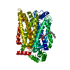 4zw9S S: Starting model for refinement C: citing same article ( |
|---|---|
| Similar structure data |
-Links
 PDBj
PDBj


- Assembly
| Deposited unit | 
| ||||||||
|---|---|---|---|---|---|---|---|---|---|
| 1 | 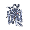
| ||||||||
| 2 | 
| ||||||||
| Unit cell |
|
-Components
| #1: Protein | Mass: 56907.918 Da / Num. of mol.: 2 / Mutation: N43T Source method: isolated from a genetically manipulated source Source: (gene. exp.) Homo sapiens (human) / Gene: SLC2A3, GLUT3 / Production host:   Spodoptera frugiperda (fall armyworm) / References: UniProt: P11169 Spodoptera frugiperda (fall armyworm) / References: UniProt: P11169#2: Chemical |   Mass: 519.571 Da / Num. of mol.: 2 / Source method: obtained synthetically / Formula: C27H30FN7O3 / Feature type: SUBJECT OF INVESTIGATION Mass: 519.571 Da / Num. of mol.: 2 / Source method: obtained synthetically / Formula: C27H30FN7O3 / Feature type: SUBJECT OF INVESTIGATION#3: Chemical | ChemComp-OLC / (   Mass: 356.540 Da / Num. of mol.: 5 / Source method: obtained synthetically / Formula: C21H40O4 Mass: 356.540 Da / Num. of mol.: 5 / Source method: obtained synthetically / Formula: C21H40O4#4: Water | ChemComp-HOH / |  Mass: 18.015 Da / Num. of mol.: 118 / Source method: isolated from a natural source / Formula: H2O Mass: 18.015 Da / Num. of mol.: 118 / Source method: isolated from a natural source / Formula: H2OHas ligand of interest | Y | |
|---|
-Experimental details
-Experiment
| Experiment | Method: X-RAY DIFFRACTION / Number of used crystals: 1 |
|---|
- Sample preparation
| Crystal | Density Matthews: 3.77 Å3/Da / Density % sol: 67.42 % |
|---|---|
| Crystal grow | Temperature: 293 K / Method: lipidic cubic phase Details: 40% v/v PEG400, 100 mM sodium citrate, pH 5.7, 400 mM potassium formate |
-Data collection
| Diffraction | Mean temperature: 80 K / Serial crystal experiment: N |
|---|---|
| Diffraction source | Source: SYNCHROTRON / Site: SPring-8  / Beamline: BL32XU / Wavelength: 1 Å / Beamline: BL32XU / Wavelength: 1 Å |
| Detector | Type: DECTRIS EIGER X 9M / Detector: PIXEL / Date: Oct 30, 2019 |
| Radiation | Protocol: SINGLE WAVELENGTH / Monochromatic (M) / Laue (L): M / Scattering type: x-ray |
| Radiation wavelength | Wavelength: 1 Å / Relative weight: 1 |
| Reflection | Resolution: 2.3→50 Å / Num. obs: 75008 / % possible obs: 99.9 % / Redundancy: 23.07 % / CC1/2: 0.998 / Net I/σ(I): 11.75 |
| Reflection shell | Resolution: 2.3→2.4 Å / Num. unique obs: 8977 / CC1/2: 0.536 |
- Processing
| Software |
| |||||||||||||||||||||||||||||||||||||||||||||||||||||||||||||||||||||||||||||||||||||||||||||||||||||||||||||||||||||||||||||||||||||||||||||||||||||||||||||||||||||||||||||||||||||||||||||||||||||||||||||||||||||||||
|---|---|---|---|---|---|---|---|---|---|---|---|---|---|---|---|---|---|---|---|---|---|---|---|---|---|---|---|---|---|---|---|---|---|---|---|---|---|---|---|---|---|---|---|---|---|---|---|---|---|---|---|---|---|---|---|---|---|---|---|---|---|---|---|---|---|---|---|---|---|---|---|---|---|---|---|---|---|---|---|---|---|---|---|---|---|---|---|---|---|---|---|---|---|---|---|---|---|---|---|---|---|---|---|---|---|---|---|---|---|---|---|---|---|---|---|---|---|---|---|---|---|---|---|---|---|---|---|---|---|---|---|---|---|---|---|---|---|---|---|---|---|---|---|---|---|---|---|---|---|---|---|---|---|---|---|---|---|---|---|---|---|---|---|---|---|---|---|---|---|---|---|---|---|---|---|---|---|---|---|---|---|---|---|---|---|---|---|---|---|---|---|---|---|---|---|---|---|---|---|---|---|---|---|---|---|---|---|---|---|---|---|---|---|---|---|---|---|---|
| Refinement | Method to determine structure: MOLECULAR REPLACEMENT Starting model: PDB entry 4ZW9 Resolution: 2.3→46.39 Å / SU ML: 0.3 / Cross valid method: THROUGHOUT / σ(F): 0.8 / Phase error: 25.55 / Stereochemistry target values: ML
| |||||||||||||||||||||||||||||||||||||||||||||||||||||||||||||||||||||||||||||||||||||||||||||||||||||||||||||||||||||||||||||||||||||||||||||||||||||||||||||||||||||||||||||||||||||||||||||||||||||||||||||||||||||||||
| Solvent computation | Shrinkage radii: 0.9 Å / VDW probe radii: 1.11 Å / Solvent model: FLAT BULK SOLVENT MODEL | |||||||||||||||||||||||||||||||||||||||||||||||||||||||||||||||||||||||||||||||||||||||||||||||||||||||||||||||||||||||||||||||||||||||||||||||||||||||||||||||||||||||||||||||||||||||||||||||||||||||||||||||||||||||||
| Displacement parameters | Biso max: 159.41 Å2 / Biso mean: 54.6828 Å2 / Biso min: 26.9 Å2 | |||||||||||||||||||||||||||||||||||||||||||||||||||||||||||||||||||||||||||||||||||||||||||||||||||||||||||||||||||||||||||||||||||||||||||||||||||||||||||||||||||||||||||||||||||||||||||||||||||||||||||||||||||||||||
| Refinement step | Cycle: final / Resolution: 2.3→46.39 Å
| |||||||||||||||||||||||||||||||||||||||||||||||||||||||||||||||||||||||||||||||||||||||||||||||||||||||||||||||||||||||||||||||||||||||||||||||||||||||||||||||||||||||||||||||||||||||||||||||||||||||||||||||||||||||||
| LS refinement shell | Refine-ID: X-RAY DIFFRACTION / Rfactor Rfree error: 0 / Total num. of bins used: 30
|
