 Movie
Movie Controller


+ Open data

- Basic information
| Entry | Database: PDB / ID: 7snd | ||||||
|---|---|---|---|---|---|---|---|
| Title | Pacifastin related protease inhibitors | ||||||
 Components Components | Pacifastin-related peptide | ||||||
 Keywords Keywords | TOXIN / CDP / Pacifastin / protease inhibitor | ||||||
| Function / homology | Pacifastin domain / Pacifastin domain superfamily / Pacifastin inhibitor (LCMII) / Pacifastin domain profile. / serine-type endopeptidase inhibitor activity / extracellular region / PHOSPHATE ION / Pacifastin-related peptide Function and homology information Function and homology information | ||||||
| Biological species |  | ||||||
| Method |  X-RAY DIFFRACTION / MOLECULAR REPLACEMENT / Resolution: 1.79 Å X-RAY DIFFRACTION / MOLECULAR REPLACEMENT / Resolution: 1.79 Å | ||||||
 Authors Authors | Gewe, M.M. / Strong, R.K. | ||||||
| Funding support | 1items
| ||||||
 Citation Citation | Journal: Sci Transl Med / Year: 2022 Title: Ex silico engineering of cystine-dense peptides yielding a potent bispecific T cell engager. Authors: Crook, Z.R. / Girard, E.J. / Sevilla, G.P. / Brusniak, M.Y. / Rupert, P.B. / Friend, D.J. / Gewe, M.M. / Clarke, M. / Lin, I. / Ruff, R. / Pakiam, F. / Phi, T.D. / Bandaranayake, A. / ...Authors: Crook, Z.R. / Girard, E.J. / Sevilla, G.P. / Brusniak, M.Y. / Rupert, P.B. / Friend, D.J. / Gewe, M.M. / Clarke, M. / Lin, I. / Ruff, R. / Pakiam, F. / Phi, T.D. / Bandaranayake, A. / Correnti, C.E. / Mhyre, A.J. / Nairn, N.W. / Strong, R.K. / Olson, J.M. #1: Journal: Nat Struct Mol Biol / Year: 2018Title: Screening, large-scale production and structure-based classification of cystine-dense peptides Authors: Correnti, C.E. / Gewe, M.M. / Mehlin, C. / Bandaranayake, A.D. / Johnsen, W.A. / Rupert, P.B. / Brusniak, M.Y. / Clarke, M. / Burke, S.E. / De Van Der Schueren, W. / Pilat, K. / Turnbaugh, S. ...Authors: Correnti, C.E. / Gewe, M.M. / Mehlin, C. / Bandaranayake, A.D. / Johnsen, W.A. / Rupert, P.B. / Brusniak, M.Y. / Clarke, M. / Burke, S.E. / De Van Der Schueren, W. / Pilat, K. / Turnbaugh, S.M. / May, D. / Watson, A. / Chan, M.K. / Bahl, C.D. / Olson, J.M. / Strong, R.K. | ||||||
| History |
|
- Structure visualization
| Structure viewer | Molecule: MolmilJmol/JSmol |
|---|
- Downloads & links
-Download
| PDBx/mmCIF format | 7snd.cif.gz | 40.4 KB | Display | PDBx/mmCIF format |
|---|---|---|---|---|
| PDB format | pdb7snd.ent.gz | 27 KB | Display | PDB format |
| PDBx/mmJSON format | 7snd.json.gz | Tree view | PDBx/mmJSON format | |
| Others |  Other downloads Other downloads |
-Validation report
| Arichive directory | https://data.pdbj.org/pub/pdb/validation_reports/sn/7sndftp://data.pdbj.org/pub/pdb/validation_reports/sn/7snd | HTTPS FTP |
|---|
-Related structure data
| Related structure data | 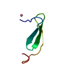 7saoC  7sapC  7sgqC  7sjqC  7sltC  7sncC  1gl1S S: Starting model for refinement C: citing same article ( |
|---|---|
| Similar structure data |
-Links
 PDBj
PDBj- Assembly
| Deposited unit | 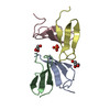
| ||||||||
|---|---|---|---|---|---|---|---|---|---|
| 1 | 
| ||||||||
| 2 | 
| ||||||||
| 3 | 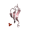
| ||||||||
| 4 | 
| ||||||||
| Unit cell |
|
-Components
| #1: Protein/peptide | Mass: 3558.979 Da / Num. of mol.: 4 Source method: isolated from a genetically manipulated source Source: (gene. exp.) Production host:  Homo sapiens (human) / References: UniProt: R4UK43 Homo sapiens (human) / References: UniProt: R4UK43#2: Chemical |   Mass: 92.094 Da / Num. of mol.: 2 / Source method: obtained synthetically / Formula: C3H8O3 / Feature type: SUBJECT OF INVESTIGATION Mass: 92.094 Da / Num. of mol.: 2 / Source method: obtained synthetically / Formula: C3H8O3 / Feature type: SUBJECT OF INVESTIGATION#3: Chemical | ChemComp-PO4 / |   Mass: 94.971 Da / Num. of mol.: 1 / Source method: isolated from a natural source / Formula: PO4 / Feature type: SUBJECT OF INVESTIGATION Mass: 94.971 Da / Num. of mol.: 1 / Source method: isolated from a natural source / Formula: PO4 / Feature type: SUBJECT OF INVESTIGATION#4: Water | ChemComp-HOH / |  Mass: 18.015 Da / Num. of mol.: 88 / Source method: isolated from a natural source / Formula: H2O Mass: 18.015 Da / Num. of mol.: 88 / Source method: isolated from a natural source / Formula: H2OHas ligand of interest | Y | Has protein modification | Y | |
|---|
-Experimental details
-Experiment
| Experiment | Method: X-RAY DIFFRACTION / Number of used crystals: 1 |
|---|
- Sample preparation
| Crystal | Density Matthews: 1.65 Å3/Da / Density % sol: 25.36 % |
|---|---|
| Crystal grow | Temperature: 298 K / Method: vapor diffusion, sitting drop Details: 0.2 M Lithium Chloride, 0.1 M Phosphate-Citrate pH 4.2, 20% PEG 1000 |
-Data collection
| Diffraction | Mean temperature: 100 K / Serial crystal experiment: N | |||||||||||||||||||||||||||||||||||||||||||||||||||||||||||||||||||||||||||||||||||||||||||||||||||||||||||||||||||||||||||||||||||||||||||||||||||||||||||||||||||||||||||||||||||||||||||||
|---|---|---|---|---|---|---|---|---|---|---|---|---|---|---|---|---|---|---|---|---|---|---|---|---|---|---|---|---|---|---|---|---|---|---|---|---|---|---|---|---|---|---|---|---|---|---|---|---|---|---|---|---|---|---|---|---|---|---|---|---|---|---|---|---|---|---|---|---|---|---|---|---|---|---|---|---|---|---|---|---|---|---|---|---|---|---|---|---|---|---|---|---|---|---|---|---|---|---|---|---|---|---|---|---|---|---|---|---|---|---|---|---|---|---|---|---|---|---|---|---|---|---|---|---|---|---|---|---|---|---|---|---|---|---|---|---|---|---|---|---|---|---|---|---|---|---|---|---|---|---|---|---|---|---|---|---|---|---|---|---|---|---|---|---|---|---|---|---|---|---|---|---|---|---|---|---|---|---|---|---|---|---|---|---|---|---|---|---|---|---|
| Diffraction source | Source: ROTATING ANODE / Type: RIGAKU MICROMAX-007 HF / Wavelength: 1.54 Å | |||||||||||||||||||||||||||||||||||||||||||||||||||||||||||||||||||||||||||||||||||||||||||||||||||||||||||||||||||||||||||||||||||||||||||||||||||||||||||||||||||||||||||||||||||||||||||||
| Detector | Type: RIGAKU SATURN 944+ / Detector: CCD / Date: Oct 7, 2016 | |||||||||||||||||||||||||||||||||||||||||||||||||||||||||||||||||||||||||||||||||||||||||||||||||||||||||||||||||||||||||||||||||||||||||||||||||||||||||||||||||||||||||||||||||||||||||||||
| Radiation | Protocol: SINGLE WAVELENGTH / Monochromatic (M) / Laue (L): M / Scattering type: x-ray | |||||||||||||||||||||||||||||||||||||||||||||||||||||||||||||||||||||||||||||||||||||||||||||||||||||||||||||||||||||||||||||||||||||||||||||||||||||||||||||||||||||||||||||||||||||||||||||
| Radiation wavelength | Wavelength: 1.54 Å / Relative weight: 1 | |||||||||||||||||||||||||||||||||||||||||||||||||||||||||||||||||||||||||||||||||||||||||||||||||||||||||||||||||||||||||||||||||||||||||||||||||||||||||||||||||||||||||||||||||||||||||||||
| Reflection | Resolution: 1.79→50 Å / Num. obs: 6336 / % possible obs: 72.2 % / Redundancy: 7.2 % / Rmerge(I) obs: 0.068 / Rpim(I) all: 0.027 / Rrim(I) all: 0.073 / Χ2: 2.337 / Net I/σ(I): 17.1 / Num. measured all: 45734 | |||||||||||||||||||||||||||||||||||||||||||||||||||||||||||||||||||||||||||||||||||||||||||||||||||||||||||||||||||||||||||||||||||||||||||||||||||||||||||||||||||||||||||||||||||||||||||||
| Reflection shell | Diffraction-ID: 1
|
- Processing
| Software |
| ||||||||||||||||||||||||||||||||||||||||||||||||||||||||||||
|---|---|---|---|---|---|---|---|---|---|---|---|---|---|---|---|---|---|---|---|---|---|---|---|---|---|---|---|---|---|---|---|---|---|---|---|---|---|---|---|---|---|---|---|---|---|---|---|---|---|---|---|---|---|---|---|---|---|---|---|---|---|
| Refinement | Method to determine structure: MOLECULAR REPLACEMENT Starting model: 1GL1 Resolution: 1.79→28.75 Å / Cor.coef. Fo:Fc: 0.966 / Cor.coef. Fo:Fc free: 0.932 / SU B: 2.5 / SU ML: 0.078 / Cross valid method: THROUGHOUT / σ(F): 0 / ESU R: 0.227 / ESU R Free: 0.179 / Stereochemistry target values: MAXIMUM LIKELIHOOD Details: HYDROGENS HAVE BEEN ADDED IN THE RIDING POSITIONS U VALUES : REFINED INDIVIDUALLY
| ||||||||||||||||||||||||||||||||||||||||||||||||||||||||||||
| Solvent computation | Ion probe radii: 0.8 Å / Shrinkage radii: 0.8 Å / VDW probe radii: 1.2 Å / Solvent model: MASK | ||||||||||||||||||||||||||||||||||||||||||||||||||||||||||||
| Displacement parameters | Biso max: 58.99 Å2 / Biso mean: 16.267 Å2 / Biso min: 9.04 Å2
| ||||||||||||||||||||||||||||||||||||||||||||||||||||||||||||
| Refinement step | Cycle: final / Resolution: 1.79→28.75 Å
| ||||||||||||||||||||||||||||||||||||||||||||||||||||||||||||
| Refine LS restraints |
| ||||||||||||||||||||||||||||||||||||||||||||||||||||||||||||
| LS refinement shell | Resolution: 1.791→1.837 Å / Rfactor Rfree error: 0 / Total num. of bins used: 20
|
