 Movie
Movie Controller

[English] 日本語
 Yorodumi
- PDB-7sk9: Cryo-EM structure of human ACKR3 in complex with a small molecule... -
+ Open data

- Basic information
| Entry | Database: PDB / ID: 7sk9 | |||||||||||||||||||||||||||
|---|---|---|---|---|---|---|---|---|---|---|---|---|---|---|---|---|---|---|---|---|---|---|---|---|---|---|---|---|
| Title | Cryo-EM structure of human ACKR3 in complex with a small molecule partial agonist CCX662, and an intracellular Fab | |||||||||||||||||||||||||||
 Components Components |
| |||||||||||||||||||||||||||
 Keywords Keywords | SIGNALING PROTEIN/IMMUNE SYSTEM / Atypical Chemokine Receptor / MEMBRANE PROTEIN / SIGNALING PROTEIN / SIGNALING PROTEIN-IMMUNE SYSTEM complex | |||||||||||||||||||||||||||
| Function / homology |  Function and homology information Function and homology informationoculomotor nerve development / positive regulation of mesenchymal stem cell migration / C-X-C chemokine binding / C-X-C chemokine receptor activity / negative regulation of intrinsic apoptotic signaling pathway in response to DNA damage / C-C chemokine receptor activity / C-C chemokine binding / scavenger receptor activity / Chemokine receptors bind chemokines / vasculogenesis ...oculomotor nerve development / positive regulation of mesenchymal stem cell migration / C-X-C chemokine binding / C-X-C chemokine receptor activity / negative regulation of intrinsic apoptotic signaling pathway in response to DNA damage / C-C chemokine receptor activity / C-C chemokine binding / scavenger receptor activity / Chemokine receptors bind chemokines / vasculogenesis / coreceptor activity / clathrin-coated pit / chemokine-mediated signaling pathway / cell chemotaxis / calcium-mediated signaling / receptor internalization / recycling endosome / positive regulation of cytosolic calcium ion concentration / angiogenesis / G alpha (i) signalling events / early endosome / positive regulation of ERK1 and ERK2 cascade / cell adhesion / endosome / immune response / external side of plasma membrane / negative regulation of cell population proliferation / cell surface / plasma membrane Similarity search - Function | |||||||||||||||||||||||||||
| Biological species |  Homo sapiens (human) Homo sapiens (human) | |||||||||||||||||||||||||||
| Method | ELECTRON MICROSCOPY / single particle reconstruction / cryo EM / Resolution: 3.7 Å | |||||||||||||||||||||||||||
 Authors Authors | Yen, Y.C. / Schafer, C.T. / Gustavsson, M. / Handel, T.M. / Tesmer, J.J.G. | |||||||||||||||||||||||||||
| Funding support |  United States, 7items United States, 7items
| |||||||||||||||||||||||||||
 Citation Citation | Journal: Sci Adv / Year: 2022 Title: Structures of atypical chemokine receptor 3 reveal the basis for its promiscuity and signaling bias. Authors: Yu-Chen Yen / Christopher T Schafer / Martin Gustavsson / Stefanie A Eberle / Pawel K Dominik / Dawid Deneka / Penglie Zhang / Thomas J Schall / Anthony A Kossiakoff / John J G Tesmer / Tracy M Handel /   Abstract: Both CXC chemokine receptor 4 (CXCR4) and atypical chemokine receptor 3 (ACKR3) are activated by the chemokine CXCL12 yet evoke distinct cellular responses. CXCR4 is a canonical G protein-coupled ...Both CXC chemokine receptor 4 (CXCR4) and atypical chemokine receptor 3 (ACKR3) are activated by the chemokine CXCL12 yet evoke distinct cellular responses. CXCR4 is a canonical G protein-coupled receptor (GPCR), whereas ACKR3 is intrinsically biased for arrestin. The molecular basis for this difference is not understood. Here, we describe cryo-EM structures of ACKR3 in complex with CXCL12, a more potent CXCL12 variant, and a small-molecule agonist. The bound chemokines adopt an unexpected pose relative to those established for CXCR4 and observed in other receptor-chemokine complexes. Along with functional studies, these structures provide insight into the ligand-binding promiscuity of ACKR3, why it fails to couple to G proteins, and its bias toward β-arrestin. The results lay the groundwork for understanding the physiological interplay of ACKR3 with other GPCRs. | |||||||||||||||||||||||||||
| History |
|
- Structure visualization
| Structure viewer | Molecule: MolmilJmol/JSmol |
|---|
- Downloads & links
-Download
| PDBx/mmCIF format | 7sk9.cif.gz | 251.3 KB | Display | PDBx/mmCIF format |
|---|---|---|---|---|
| PDB format | pdb7sk9.ent.gz | 201.9 KB | Display | PDB format |
| PDBx/mmJSON format | 7sk9.json.gz | Tree view | PDBx/mmJSON format | |
| Others |  Other downloads Other downloads |
-Validation report
| Arichive directory | https://data.pdbj.org/pub/pdb/validation_reports/sk/7sk9ftp://data.pdbj.org/pub/pdb/validation_reports/sk/7sk9 | HTTPS FTP |
|---|
-Related structure data
| Related structure data |  25177MC 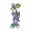 7sk3C 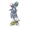 7sk4C 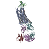 7sk5C 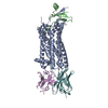 7sk6C 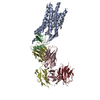 7sk7C 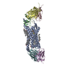 7sk8C M: map data used to model this data C: citing same article ( |
|---|---|
| Similar structure data |


-Links
 PDBj
PDBj


- Assembly
| Deposited unit | 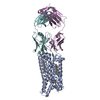
|
|---|---|
| 1 |
|
-Components
| #1: Protein | Mass: 45196.406 Da / Num. of mol.: 1 Source method: isolated from a genetically manipulated source Source: (gene. exp.) Homo sapiens (human) / Gene: ACKR3, CMKOR1, CXCR7, GPR159, RDC1 / Production host:   Spodoptera frugiperda (fall armyworm) / References: UniProt: P25106 Spodoptera frugiperda (fall armyworm) / References: UniProt: P25106 | ||||
|---|---|---|---|---|---|
| #2: Antibody | Mass: 25380.223 Da / Num. of mol.: 1 Source method: isolated from a genetically manipulated source Source: (gene. exp.) Homo sapiens (human)Production host:  | ||||
| #3: Antibody | Mass: 23471.031 Da / Num. of mol.: 1 Source method: isolated from a genetically manipulated source Source: (gene. exp.) Homo sapiens (human)Production host: | ||||
| #4: Chemical | ChemComp-GJ9 / (  Mass: 540.697 Da / Num. of mol.: 1 / Source method: obtained synthetically / Formula: C28H38N5O4S / Feature type: SUBJECT OF INVESTIGATION Mass: 540.697 Da / Num. of mol.: 1 / Source method: obtained synthetically / Formula: C28H38N5O4S / Feature type: SUBJECT OF INVESTIGATION | ||||
| #5: Chemical |   Mass: 386.654 Da / Num. of mol.: 2 / Source method: obtained synthetically / Formula: C27H46O / Feature type: SUBJECT OF INVESTIGATION Mass: 386.654 Da / Num. of mol.: 2 / Source method: obtained synthetically / Formula: C27H46O / Feature type: SUBJECT OF INVESTIGATIONHas ligand of interest | Y | Has protein modification | Y | |
-Experimental details
-Experiment
| Experiment | Method: ELECTRON MICROSCOPY |
|---|---|
| EM experiment | Aggregation state: PARTICLE / 3D reconstruction method: single particle reconstruction |
- Sample preparation
| Component | Name: Complex structure of ACKR3-CCX662- CID24 / Type: COMPLEX / Entity ID: #1-#3 / Source: MULTIPLE SOURCES | |||||||||||||||||||||||||
|---|---|---|---|---|---|---|---|---|---|---|---|---|---|---|---|---|---|---|---|---|---|---|---|---|---|---|
| Molecular weight | Experimental value: NO | |||||||||||||||||||||||||
| Buffer solution | pH: 8 | |||||||||||||||||||||||||
| Buffer component |
| |||||||||||||||||||||||||
| Specimen | Conc.: 0.2 mg/ml / Embedding applied: NO / Shadowing applied: NO / Staining applied: NO / Vitrification applied: YES | |||||||||||||||||||||||||
| Specimen support | Grid material: COPPER / Grid mesh size: 300 divisions/in. / Grid type: Quantifoil R1.2/1.3 | |||||||||||||||||||||||||
| Vitrification | Instrument: FEI VITROBOT MARK IV / Cryogen name: ETHANE / Humidity: 100 % / Chamber temperature: 277 K |
- Electron microscopy imaging
| Experimental equipment |  Model: Titan Krios / Image courtesy: FEI Company |
|---|---|
| Microscopy | Model: FEI TITAN KRIOS |
| Electron gun | Electron source:  FIELD EMISSION GUN / Accelerating voltage: 300 kV / Illumination mode: FLOOD BEAM FIELD EMISSION GUN / Accelerating voltage: 300 kV / Illumination mode: FLOOD BEAM |
| Electron lens | Mode: BRIGHT FIELD / Cs: 2.7 mm / C2 aperture diameter: 100 µm |
| Specimen holder | Cryogen: NITROGEN / Specimen holder model: FEI TITAN KRIOS AUTOGRID HOLDER |
| Image recording | Electron dose: 53.8 e/Å2 / Film or detector model: GATAN K3 (6k x 4k) |
- Processing
| Software | Name: PHENIX / Version: 1.19.2_4158: / Classification: refinement | ||||||||||||||||||||||||
|---|---|---|---|---|---|---|---|---|---|---|---|---|---|---|---|---|---|---|---|---|---|---|---|---|---|
| EM software |
| ||||||||||||||||||||||||
| CTF correction | Type: PHASE FLIPPING AND AMPLITUDE CORRECTION | ||||||||||||||||||||||||
| Symmetry | Point symmetry: C1 (asymmetric) | ||||||||||||||||||||||||
| 3D reconstruction | Resolution: 3.7 Å / Resolution method: FSC 0.143 CUT-OFF / Num. of particles: 297347 / Symmetry type: POINT | ||||||||||||||||||||||||
| Refine LS restraints |
|
