Movie
Movie Controller

[English] 日本語
 Yorodumi
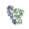
- PDB-7sjx: Cryo-EM Structure of the PR-RT components of the HIV-1 Pol Polyprotein -
+ Open data

- Basic information
| Entry | Database: PDB / ID: 7sjx | ||||||||||||
|---|---|---|---|---|---|---|---|---|---|---|---|---|---|
| Title | Cryo-EM Structure of the PR-RT components of the HIV-1 Pol Polyprotein | ||||||||||||
 Components Components | Gag-Pol polyprotein | ||||||||||||
 Keywords Keywords | VIRAL PROTEIN / HIV-1 / Reverse transcriptase / Protease / enzyme | ||||||||||||
| Function / homology |  Function and homology information Function and homology informationHIV-1 retropepsin / symbiont-mediated activation of host apoptosis / retroviral ribonuclease H / exoribonuclease H / exoribonuclease H activity / DNA integration / viral genome integration into host DNA / establishment of integrated proviral latency / RNA-directed DNA polymerase / RNA stem-loop binding ...HIV-1 retropepsin / symbiont-mediated activation of host apoptosis / retroviral ribonuclease H / exoribonuclease H / exoribonuclease H activity / DNA integration / viral genome integration into host DNA / establishment of integrated proviral latency / RNA-directed DNA polymerase / RNA stem-loop binding / viral penetration into host nucleus / host multivesicular body / RNA-directed DNA polymerase activity / RNA-DNA hybrid ribonuclease activity / Transferases; Transferring phosphorus-containing groups; Nucleotidyltransferases / host cell / viral nucleocapsid / DNA recombination / DNA-directed DNA polymerase / aspartic-type endopeptidase activity / Hydrolases; Acting on ester bonds / DNA-directed DNA polymerase activity / symbiont-mediated suppression of host gene expression / viral translational frameshifting / symbiont entry into host cell / lipid binding / host cell nucleus / host cell plasma membrane / virion membrane / structural molecule activity / proteolysis / DNA binding / zinc ion binding Similarity search - Function | ||||||||||||
| Biological species |  Human immunodeficiency virus type 1 group M subtype B Human immunodeficiency virus type 1 group M subtype B | ||||||||||||
| Method | ELECTRON MICROSCOPY / single particle reconstruction / cryo EM / Resolution: 8.2 Å | ||||||||||||
 Authors Authors | Lyumkis, D. / Passos, D. / Arnold, E. / Harrison, J.J.E.K. / Ruiz, F.X. | ||||||||||||
| Funding support |  United States, 3items United States, 3items
| ||||||||||||
 Citation Citation | Journal: Sci Adv / Year: 2022 Title: Cryo-EM structure of the HIV-1 Pol polyprotein provides insights into virion maturation. Authors: Jerry Joe E K Harrison / Dario Oliveira Passos / Jessica F Bruhn / Joseph D Bauman / Lynda Tuberty / Jeffrey J DeStefano / Francesc Xavier Ruiz / Dmitry Lyumkis / Eddy Arnold / Abstract: Key proteins of retroviruses and other RNA viruses are translated and subsequently processed from polyprotein precursors by the viral protease (PR). Processing of the HIV Gag-Pol polyprotein yields ...Key proteins of retroviruses and other RNA viruses are translated and subsequently processed from polyprotein precursors by the viral protease (PR). Processing of the HIV Gag-Pol polyprotein yields the HIV structural proteins and enzymes. Structures of the mature enzymes PR, reverse transcriptase (RT), and integrase (IN) aided understanding of catalysis and design of antiretrovirals, but knowledge of the Pol precursor architecture and function before PR cleavage is limited. We developed a system to produce stable HIV-1 Pol and determined its cryo-electron microscopy structure. RT in Pol has a similar arrangement to the mature RT heterodimer, and its dimerization may draw together two PR monomers to activate proteolytic processing. HIV-1 thus may leverage the dimerization interfaces in Pol to regulate assembly and maturation of polyprotein precursors. | ||||||||||||
| History |
|
- Structure visualization
| Structure viewer | Molecule: MolmilJmol/JSmol |
|---|
- Downloads & links
-Download
| PDBx/mmCIF format | 7sjx.cif.gz | 249.9 KB | Display | PDBx/mmCIF format |
|---|---|---|---|---|
| PDB format | pdb7sjx.ent.gz | Display | PDB format | |
| PDBx/mmJSON format | 7sjx.json.gz | Tree view | PDBx/mmJSON format | |
| Others |  Other downloads Other downloads |
-Validation report
| Arichive directory | https://data.pdbj.org/pub/pdb/validation_reports/sj/7sjxftp://data.pdbj.org/pub/pdb/validation_reports/sj/7sjx | HTTPS FTP |
|---|
-Related structure data
| Related structure data |  25165MC 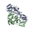 7sepC M: map data used to model this data C: citing same article ( |
|---|---|
| Similar structure data |


-Links
 PDBj
PDBj


- Assembly
| Deposited unit | 
|
|---|---|
| 1 |
|
-Components
| #1: Protein | Mass: 119289.867 Da / Num. of mol.: 2 / Fragment: PR-RT portion, residues 479-1447 / Mutation: D131A, L765D, F766D Source method: isolated from a genetically manipulated source Source: (gene. exp.) Human immunodeficiency virus type 1 group M subtype B (isolate BH10)Strain: isolate BH10 / Gene: gag-pol / Production host:  |
|---|
-Experimental details
-Experiment
| Experiment | Method: ELECTRON MICROSCOPY |
|---|---|
| EM experiment | Aggregation state: PARTICLE / 3D reconstruction method: single particle reconstruction |
- Sample preparation
| Component | Name: PR-RT portion of HIV-1 Pol / Type: COMPLEX Details: 3D reconstruction of the HIV-1 Pol polyprotein comprising the PR-RT portion Entity ID: all / Source: RECOMBINANT |
|---|---|
| Molecular weight | Value: 0.13 MDa / Experimental value: NO |
| Source (natural) | Organism: Human immunodeficiency virus type 1 BH10 |
| Source (recombinant) | Organism: |
| Buffer solution | pH: 8 |
| Specimen | Conc.: 0.3 mg/ml / Embedding applied: NO / Shadowing applied: NO / Staining applied: NO / Vitrification applied: YES |
| Vitrification | Cryogen name: ETHANE |
- Electron microscopy imaging
| Experimental equipment |  Model: Titan Krios / Image courtesy: FEI Company |
|---|---|
| Microscopy | Model: FEI TITAN KRIOS |
| Electron gun | Electron source:  FIELD EMISSION GUN / Accelerating voltage: 300 kV / Illumination mode: FLOOD BEAM FIELD EMISSION GUN / Accelerating voltage: 300 kV / Illumination mode: FLOOD BEAM |
| Electron lens | Mode: BRIGHT FIELD / Cs: 2.7 mm |
| Image recording | Electron dose: 0.95 e/Å2 / Detector mode: COUNTING / Film or detector model: GATAN K2 SUMMIT (4k x 4k) |
| Image scans | Width: 3710 / Height: 3838 / Movie frames/image: 60 |
- Processing
| Software |
| ||||||||||||||||||||||||||||||||||||||||||||
|---|---|---|---|---|---|---|---|---|---|---|---|---|---|---|---|---|---|---|---|---|---|---|---|---|---|---|---|---|---|---|---|---|---|---|---|---|---|---|---|---|---|---|---|---|---|
| EM software |
| ||||||||||||||||||||||||||||||||||||||||||||
| CTF correction | Type: PHASE FLIPPING AND AMPLITUDE CORRECTION | ||||||||||||||||||||||||||||||||||||||||||||
| Particle selection | Num. of particles selected: 27555 | ||||||||||||||||||||||||||||||||||||||||||||
| 3D reconstruction | Resolution: 8.2 Å / Resolution method: FSC 0.143 CUT-OFF / Num. of particles: 27555 / Symmetry type: POINT | ||||||||||||||||||||||||||||||||||||||||||||
| Atomic model building | Protocol: RIGID BODY FIT / Space: REAL / Target criteria: Correlation coeficient | ||||||||||||||||||||||||||||||||||||||||||||
| Atomic model building |
| ||||||||||||||||||||||||||||||||||||||||||||
| Refinement | Cross valid method: NONE Stereochemistry target values: GeoStd + Monomer Library + CDL v1.2 | ||||||||||||||||||||||||||||||||||||||||||||
| Displacement parameters | Biso mean: 526.76 Å2 | ||||||||||||||||||||||||||||||||||||||||||||
| Refine LS restraints |
|