 Movie
Movie Controller


+ Open data

- Basic information
| Entry | Database: PDB / ID: 7shl | ||||||||||||
|---|---|---|---|---|---|---|---|---|---|---|---|---|---|
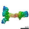
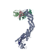
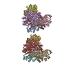
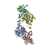
| Title | Structure of Xenopus laevis CRL2Lrr1 (State 2) | ||||||||||||
 Components Components |
| ||||||||||||
 Keywords Keywords | LIGASE / Cullin RING E3 ubiquitin ligase / DNA replication termination | ||||||||||||
| Function / homology |  Function and homology information Function and homology informationcullin-RING ubiquitin ligase complex / elongin complex / VCB complex / ubiquitin ligase complex / transcription elongation by RNA polymerase II / ubiquitin-dependent protein catabolic process / protein-macromolecule adaptor activity / protein ubiquitination / ubiquitin protein ligase binding / zinc ion binding / cytoplasm Similarity search - Function | ||||||||||||
| Biological species | |||||||||||||
| Method | ELECTRON MICROSCOPY / single particle reconstruction / cryo EM / Resolution: 3.5 Å | ||||||||||||
 Authors Authors | Zhou, H. / Brown, A. | ||||||||||||
| Funding support |  United States, 3items United States, 3items
| ||||||||||||
 Citation Citation | Journal: Nucleic Acids Res / Year: 2021 Title: Structure of CRL2Lrr1, the E3 ubiquitin ligase that promotes DNA replication termination in vertebrates. Authors: Haixia Zhou / Manal S Zaher / Johannes C Walter / Alan Brown / Abstract: When vertebrate replisomes from neighboring origins converge, the Mcm7 subunit of the replicative helicase, CMG, is ubiquitylated by the E3 ubiquitin ligase, CRL2Lrr1. Polyubiquitylated CMG is then ...When vertebrate replisomes from neighboring origins converge, the Mcm7 subunit of the replicative helicase, CMG, is ubiquitylated by the E3 ubiquitin ligase, CRL2Lrr1. Polyubiquitylated CMG is then disassembled by the p97 ATPase, leading to replication termination. To avoid premature replisome disassembly, CRL2Lrr1 is only recruited to CMGs after they converge, but the underlying mechanism is unclear. Here, we use cryogenic electron microscopy to determine structures of recombinant Xenopus laevis CRL2Lrr1 with and without neddylation. The structures reveal that CRL2Lrr1 adopts an unusually open architecture, in which the putative substrate-recognition subunit, Lrr1, is located far from the catalytic module that catalyzes ubiquitin transfer. We further demonstrate that a predicted, flexible pleckstrin homology domain at the N-terminus of Lrr1 is essential to target CRL2Lrr1 to terminated CMGs. We propose a hypothetical model that explains how CRL2Lrr1's catalytic module is positioned next to the ubiquitylation site on Mcm7, and why CRL2Lrr1 binds CMG only after replisomes converge. | ||||||||||||
| History |
|
- Structure visualization
| Movie |
Movie viewer |
|---|---|
| Structure viewer | Molecule: MolmilJmol/JSmol |
- Downloads & links
-Download
| PDBx/mmCIF format | 7shl.cif.gz | 221.5 KB | Display | PDBx/mmCIF format |
|---|---|---|---|---|
| PDB format | pdb7shl.ent.gz | 171.7 KB | Display | PDB format |
| PDBx/mmJSON format | 7shl.json.gz | Tree view | PDBx/mmJSON format | |
| Others |  Other downloads Other downloads |
-Validation report
| Arichive directory | https://data.pdbj.org/pub/pdb/validation_reports/sh/7shlftp://data.pdbj.org/pub/pdb/validation_reports/sh/7shl | HTTPS FTP |
|---|
-Related structure data
| Related structure data |  25128MC  7shkC M: map data used to model this data C: citing same article ( |
|---|---|
| Similar structure data |


-Links
 PDBj
PDBj


- Assembly
| Deposited unit | 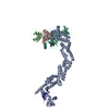
|
|---|---|
| 1 |
|
-Components
-Protein , 5 types, 5 molecules ABCLR
| #1: Protein | Mass: 87244.117 Da / Num. of mol.: 1 Source method: isolated from a genetically manipulated source Source: (gene. exp.)   Spodoptera frugiperda (fall armyworm) / References: UniProt: A0A1L8FVB6 Spodoptera frugiperda (fall armyworm) / References: UniProt: A0A1L8FVB6 |
|---|---|
| #2: Protein | Mass: 13325.927 Da / Num. of mol.: 1 Source method: isolated from a genetically manipulated source Source: (gene. exp.) Spodoptera frugiperda (fall armyworm) / References: UniProt: Q6PHL7 |
| #3: Protein | Mass: 12485.135 Da / Num. of mol.: 1 Source method: isolated from a genetically manipulated source Source: (gene. exp.) Gene: eloc.L, eloc, tceb1, XELAEV_18031836mg, XELAEV_18033803mg Production host: Spodoptera frugiperda (fall armyworm) / References: UniProt: Q6PHW7 |
| #4: Protein | Mass: 46882.258 Da / Num. of mol.: 1 Source method: isolated from a genetically manipulated source Source: (gene. exp.) Spodoptera frugiperda (fall armyworm) / References: UniProt: A4QNS4 |
| #5: Protein | Mass: 12277.985 Da / Num. of mol.: 1 Source method: isolated from a genetically manipulated source Source: (gene. exp.) Spodoptera frugiperda (fall armyworm) / References: UniProt: A0A1L8GNG7 |
-Non-polymers , 1 types, 1 molecules 
| #6: Chemical | ChemComp-ZN /  Mass: 65.409 Da / Num. of mol.: 1 / Source method: obtained synthetically / Formula: Zn Mass: 65.409 Da / Num. of mol.: 1 / Source method: obtained synthetically / Formula: Zn |
|---|
-Details
| Has ligand of interest | N |
|---|
-Experimental details
-Experiment
| Experiment | Method: ELECTRON MICROSCOPY |
|---|---|
| EM experiment | Aggregation state: PARTICLE / 3D reconstruction method: single particle reconstruction |
- Sample preparation
| Component | Name: CRL2Lrr1 / Type: COMPLEX / Entity ID: #1-#5 / Source: RECOMBINANT | ||||||||||||||||||||
|---|---|---|---|---|---|---|---|---|---|---|---|---|---|---|---|---|---|---|---|---|---|
| Molecular weight | Experimental value: NO | ||||||||||||||||||||
| Source (natural) | Organism: | ||||||||||||||||||||
| Source (recombinant) | Organism: Spodoptera frugiperda (fall armyworm) | ||||||||||||||||||||
| Buffer solution | pH: 7.6 / Details: TCEP is freshly added. | ||||||||||||||||||||
| Buffer component |
| ||||||||||||||||||||
| Specimen | Conc.: 0.8 mg/ml / Embedding applied: NO / Shadowing applied: NO / Staining applied: NO / Vitrification applied: YES | ||||||||||||||||||||
| Specimen support | Grid material: COPPER / Grid mesh size: 400 divisions/in. / Grid type: C-flat-1.2/1.3 | ||||||||||||||||||||
| Vitrification | Instrument: FEI VITROBOT MARK IV / Cryogen name: NITROGEN / Humidity: 100 % / Chamber temperature: 281.2 K / Details: Blot 6 seconds with the force 12 |
- Electron microscopy imaging
| Experimental equipment |  Model: Titan Krios / Image courtesy: FEI Company |
|---|---|
| Microscopy | Model: FEI TITAN KRIOS |
| Electron gun | Electron source:  FIELD EMISSION GUN / Accelerating voltage: 300 kV / Illumination mode: FLOOD BEAM FIELD EMISSION GUN / Accelerating voltage: 300 kV / Illumination mode: FLOOD BEAM |
| Electron lens | Mode: BRIGHT FIELD |
| Specimen holder | Specimen holder model: FEI TITAN KRIOS AUTOGRID HOLDER |
| Image recording | Electron dose: 54.5 e/Å2 / Film or detector model: GATAN K3 BIOQUANTUM (6k x 4k) |
- Processing
| Software | Name: PHENIX / Version: 1.19.2_4158: / Classification: refinement | ||||||||||||||||||||||||
|---|---|---|---|---|---|---|---|---|---|---|---|---|---|---|---|---|---|---|---|---|---|---|---|---|---|
| EM software |
| ||||||||||||||||||||||||
| CTF correction | Type: PHASE FLIPPING AND AMPLITUDE CORRECTION | ||||||||||||||||||||||||
| 3D reconstruction | Resolution: 3.5 Å / Resolution method: FSC 0.143 CUT-OFF / Num. of particles: 406755 / Symmetry type: POINT | ||||||||||||||||||||||||
| Atomic model building | Protocol: OTHER / Space: REAL / Target criteria: Correlation coefficient | ||||||||||||||||||||||||
| Refine LS restraints |
|
