 Movie
Movie Controller

[English] 日本語
 Yorodumi
- PDB-7sfp: Crystal structure of OssO, a spirocyclase involved in the biosynt... -
+ Open data

- Basic information
| Entry | Database: PDB / ID: 7sfp | |||||||||
|---|---|---|---|---|---|---|---|---|---|---|
| Title | Crystal structure of OssO, a spirocyclase involved in the biosynthesis of ossamycin | |||||||||
 Components Components | Spirocyclase | |||||||||
 Keywords Keywords | HYDROLASE / cyclase / spyrocyclase / oligomycin / calycin | |||||||||
| Function / homology | Putative spirocyclase Function and homology information Function and homology information | |||||||||
| Biological species |  Streptomyces ossamyceticus (bacteria) Streptomyces ossamyceticus (bacteria) | |||||||||
| Method |  X-RAY DIFFRACTION / SYNCHROTRON / MOLECULAR REPLACEMENT / Resolution: 2.2 Å X-RAY DIFFRACTION / SYNCHROTRON / MOLECULAR REPLACEMENT / Resolution: 2.2 Å | |||||||||
 Authors Authors | Bilyk, O. / Leadlay, P.F. / Dias, M.V.B. | |||||||||
| Funding support |  Brazil, 2items Brazil, 2items
| |||||||||
 Citation Citation | Journal: J.Am.Chem.Soc. / Year: 2022 Title: Enzyme-Catalyzed Spiroacetal Formation in Polyketide Antibiotic Biosynthesis. Authors: Bilyk, O. / Oliveira, G.S. / de Angelo, R.M. / Almeida, M.O. / Honorio, K.M. / Leeper, F.J. / Dias, M.V.B. / Leadlay, P.F. | |||||||||
| History |
|
- Structure visualization
| Structure viewer | Molecule: MolmilJmol/JSmol |
|---|
- Downloads & links
-Download
| PDBx/mmCIF format | 7sfp.cif.gz | 79.7 KB | Display | PDBx/mmCIF format |
|---|---|---|---|---|
| PDB format | pdb7sfp.ent.gz | 57.7 KB | Display | PDB format |
| PDBx/mmJSON format | 7sfp.json.gz | Tree view | PDBx/mmJSON format | |
| Others |  Other downloads Other downloads |
-Validation report
| Summary document | 7sfp_validation.pdf.gz | 433.8 KB | Display | wwPDB validaton report |
|---|---|---|---|---|
| Full document | 7sfp_full_validation.pdf.gz | 433.9 KB | Display | |
| Data in XML | 7sfp_validation.xml.gz | 7.8 KB | Display | |
| Data in CIF | 7sfp_validation.cif.gz | 9.6 KB | Display | |
| Arichive directory | https://data.pdbj.org/pub/pdb/validation_reports/sf/7sfpftp://data.pdbj.org/pub/pdb/validation_reports/sf/7sfp | HTTPS FTP |
-Related structure data
| Related structure data |  7sfnSC S: Starting model for refinement C: citing same article ( |
|---|---|
| Similar structure data |
-Links
 PDBj
PDBj- Assembly
| Deposited unit | 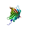
| |||||||||||||||
|---|---|---|---|---|---|---|---|---|---|---|---|---|---|---|---|---|
| 1 |
| |||||||||||||||
| Unit cell |
| |||||||||||||||
| Components on special symmetry positions |
|
-Components
| #1: Protein | Mass: 23832.330 Da / Num. of mol.: 1 Source method: isolated from a genetically manipulated source Source: (gene. exp.) Streptomyces ossamyceticus (bacteria) / Gene: ossOProduction host: References: UniProt: A0A481S8L7 |
|---|---|
| #2: Water | ChemComp-HOH /  Mass: 18.015 Da / Num. of mol.: 23 / Source method: isolated from a natural source / Formula: H2O Mass: 18.015 Da / Num. of mol.: 23 / Source method: isolated from a natural source / Formula: H2O |
-Experimental details
-Experiment
| Experiment | Method: X-RAY DIFFRACTION / Number of used crystals: 1 |
|---|
- Sample preparation
| Crystal | Density Matthews: 2.16 Å3/Da / Density % sol: 43.07 % |
|---|---|
| Crystal grow | Temperature: 293 K / Method: vapor diffusion, sitting drop / pH: 8.5 / Details: 20 %w/v PEG 8K, 0.1 M 0.2 M MgCl2 |
-Data collection
| Diffraction | Mean temperature: 100 K / Serial crystal experiment: N | |||||||||||||||||||||||||||
|---|---|---|---|---|---|---|---|---|---|---|---|---|---|---|---|---|---|---|---|---|---|---|---|---|---|---|---|---|
| Diffraction source | Source: SYNCHROTRON / Site: Diamond  / Beamline: I03 / Wavelength: 0.9795 Å / Beamline: I03 / Wavelength: 0.9795 Å | |||||||||||||||||||||||||||
| Detector | Type: DECTRIS PILATUS3 R 100K-A / Detector: PIXEL / Date: Mar 25, 2017 | |||||||||||||||||||||||||||
| Radiation | Protocol: SINGLE WAVELENGTH / Monochromatic (M) / Laue (L): M / Scattering type: x-ray | |||||||||||||||||||||||||||
| Radiation wavelength | Wavelength: 0.9795 Å / Relative weight: 1 | |||||||||||||||||||||||||||
| Reflection | Resolution: 2.2→49.02 Å / Num. obs: 11716 / % possible obs: 100 % / Redundancy: 50 % / CC1/2: 1 / Rmerge(I) obs: 0.123 / Rpim(I) all: 0.018 / Rrim(I) all: 0.124 / Net I/σ(I): 25 / Num. measured all: 586280 / Scaling rejects: 1 | |||||||||||||||||||||||||||
| Reflection shell | Diffraction-ID: 1 / % possible all: 99.9
|
- Processing
| Software |
| ||||||||||||||||||||||||||||||||||||||||
|---|---|---|---|---|---|---|---|---|---|---|---|---|---|---|---|---|---|---|---|---|---|---|---|---|---|---|---|---|---|---|---|---|---|---|---|---|---|---|---|---|---|
| Refinement | Method to determine structure: MOLECULAR REPLACEMENT Starting model: 7SFN Resolution: 2.2→46.725 Å / SU ML: 0.22 / Cross valid method: THROUGHOUT / σ(F): 1.34 / Phase error: 32.95 / Stereochemistry target values: ML
| ||||||||||||||||||||||||||||||||||||||||
| Solvent computation | Shrinkage radii: 0.9 Å / VDW probe radii: 1.11 Å / Solvent model: FLAT BULK SOLVENT MODEL | ||||||||||||||||||||||||||||||||||||||||
| Displacement parameters | Biso max: 133.38 Å2 / Biso mean: 68.077 Å2 / Biso min: 40.8 Å2 | ||||||||||||||||||||||||||||||||||||||||
| Refinement step | Cycle: final / Resolution: 2.2→46.725 Å
| ||||||||||||||||||||||||||||||||||||||||
| LS refinement shell | Refine-ID: X-RAY DIFFRACTION / Rfactor Rfree error: 0
| ||||||||||||||||||||||||||||||||||||||||
| Refinement TLS params. | Method: refined / Origin x: 37.977 Å / Origin y: 88.3981 Å / Origin z: 256.9182 Å
| ||||||||||||||||||||||||||||||||||||||||
| Refinement TLS group |
|
