 Movie
Movie Controller

[English] 日本語
 Yorodumi
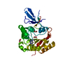
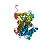
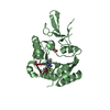
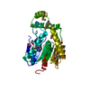
- PDB-7se8: Crystal structure of human Fibrillarin in complex with fragment 1... -
+ Open data

- Basic information
| Entry | Database: PDB / ID: 7se8 | |||||||||
|---|---|---|---|---|---|---|---|---|---|---|
| Title | Crystal structure of human Fibrillarin in complex with fragment 1 from cocktail soak | |||||||||
 Components Components | rRNA 2'-O-methyltransferase fibrillarin | |||||||||
 Keywords Keywords | TRANSFERASE / Methyltransferase / S-adenosyl methionine | |||||||||
| Function / homology |  Function and homology information Function and homology informationU6 snRNA 2'-O-ribose methyltransferase activity / snoRNA localization / granular component / histone H2AQ104 methyltransferase activity / box C/D sno(s)RNA 3'-end processing / rRNA methyltransferase activity / box C/D methylation guide snoRNP complex / rRNA methylation / rRNA modification in the nucleus and cytosol / TFIID-class transcription factor complex binding ...U6 snRNA 2'-O-ribose methyltransferase activity / snoRNA localization / granular component / histone H2AQ104 methyltransferase activity / box C/D sno(s)RNA 3'-end processing / rRNA methyltransferase activity / box C/D methylation guide snoRNP complex / rRNA methylation / rRNA modification in the nucleus and cytosol / TFIID-class transcription factor complex binding / Cajal body / Major pathway of rRNA processing in the nucleolus and cytosol / Transferases; Transferring one-carbon groups; Methyltransferases / small-subunit processome / fibrillar center / osteoblast differentiation / rRNA processing / ATPase binding / ribosomal small subunit biogenesis / nucleolus / RNA binding / extracellular exosome / nucleoplasm / membrane / nucleus Similarity search - Function | |||||||||
| Biological species |  Homo sapiens (human) Homo sapiens (human) | |||||||||
| Method |  X-RAY DIFFRACTION / SYNCHROTRON / MOLECULAR REPLACEMENT / Resolution: 1.75 Å X-RAY DIFFRACTION / SYNCHROTRON / MOLECULAR REPLACEMENT / Resolution: 1.75 Å | |||||||||
 Authors Authors | Shi, Y. / El-Deeb, I.M. / Masic, V. / Hartley-Tassell, L. / Maggioni, A. / von Itzstein, M. / Ve, T. | |||||||||
| Funding support |  Australia, 2items Australia, 2items
| |||||||||
 Citation Citation | Journal: Pharmaceuticals / Year: 2021 Title: Discovery of Cofactor Competitive Inhibitors against the Human Methyltransferase Fibrillarin. Authors: Shi, Y. / El-Deeb, I.M. / Masic, V. / Hartley-Tassell, L. / Maggioni, A. / Itzstein, M.V. / Ve, T. | |||||||||
| History |
|
- Structure visualization
| Structure viewer | Molecule: MolmilJmol/JSmol |
|---|
- Downloads & links
-Download
| PDBx/mmCIF format | 7se8.cif.gz | 231.6 KB | Display | PDBx/mmCIF format |
|---|---|---|---|---|
| PDB format | pdb7se8.ent.gz | 152.6 KB | Display | PDB format |
| PDBx/mmJSON format | 7se8.json.gz | Tree view | PDBx/mmJSON format | |
| Others |  Other downloads Other downloads |
-Validation report
| Arichive directory | https://data.pdbj.org/pub/pdb/validation_reports/se/7se8ftp://data.pdbj.org/pub/pdb/validation_reports/se/7se8 | HTTPS FTP |
|---|
-Related structure data
| Related structure data | 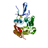 7se6C  7se7C  7se9C  7seaC  7sebC 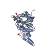 7secC 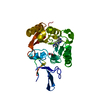 7sedC  2ipxS S: Starting model for refinement C: citing same article ( |
|---|---|
| Similar structure data |


-Links
 PDBj
PDBj


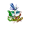
- Assembly
| Deposited unit | 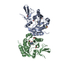
| ||||||||||||
|---|---|---|---|---|---|---|---|---|---|---|---|---|---|
| 1 | 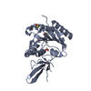
| ||||||||||||
| 2 | 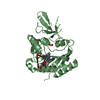
| ||||||||||||
| Unit cell |
|
-Components
| #1: Protein | Mass: 26681.590 Da / Num. of mol.: 2 Source method: isolated from a genetically manipulated source Source: (gene. exp.) Homo sapiens (human) / Gene: FBL, FIB1, FLRN / Production host:  References: UniProt: P22087, Transferases; Transferring one-carbon groups; Methyltransferases #2: Chemical | 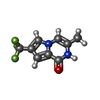  Mass: 216.160 Da / Num. of mol.: 2 / Source method: obtained synthetically / Formula: C9H7F3N2O / Feature type: SUBJECT OF INVESTIGATION Mass: 216.160 Da / Num. of mol.: 2 / Source method: obtained synthetically / Formula: C9H7F3N2O / Feature type: SUBJECT OF INVESTIGATION#3: Chemical | ChemComp-FMT /   Mass: 46.025 Da / Num. of mol.: 8 / Source method: obtained synthetically / Formula: CH2O2 Mass: 46.025 Da / Num. of mol.: 8 / Source method: obtained synthetically / Formula: CH2O2#4: Chemical |   Mass: 78.133 Da / Num. of mol.: 2 / Source method: obtained synthetically / Formula: C2H6OS / Comment: DMSO, precipitant*YM Mass: 78.133 Da / Num. of mol.: 2 / Source method: obtained synthetically / Formula: C2H6OS / Comment: DMSO, precipitant*YM#5: Water | ChemComp-HOH / |  Mass: 18.015 Da / Num. of mol.: 315 / Source method: isolated from a natural source / Formula: H2O Mass: 18.015 Da / Num. of mol.: 315 / Source method: isolated from a natural source / Formula: H2OHas ligand of interest | Y | |
|---|
-Experimental details
-Experiment
| Experiment | Method: X-RAY DIFFRACTION / Number of used crystals: 1 |
|---|
- Sample preparation
| Crystal | Density Matthews: 3.04 Å3/Da / Density % sol: 59.59 % |
|---|---|
| Crystal grow | Temperature: 293 K / Method: vapor diffusion, hanging drop / Details: 2.75-3.3 M Sodium Formate |
-Data collection
| Diffraction | Mean temperature: 100 K / Serial crystal experiment: N |
|---|---|
| Diffraction source | Source: SYNCHROTRON / Site: Australian Synchrotron / Beamline: MX1 / Wavelength: 0.9537 Å |
| Detector | Type: ADSC QUANTUM 210r / Detector: CCD / Date: Mar 7, 2018 |
| Radiation | Protocol: SINGLE WAVELENGTH / Monochromatic (M) / Laue (L): M / Scattering type: x-ray |
| Radiation wavelength | Wavelength: 0.9537 Å / Relative weight: 1 |
| Reflection | Resolution: 1.75→48.4 Å / Num. obs: 65541 / % possible obs: 98.7 % / Redundancy: 7.4 % / Biso Wilson estimate: 21.58 Å2 / CC1/2: 0.999 / Rmerge(I) obs: 0.065 / Rpim(I) all: 0.025 / Rrim(I) all: 0.07 / Net I/σ(I): 19.8 |
| Reflection shell | Resolution: 1.75→1.78 Å / Rmerge(I) obs: 0.762 / Num. unique obs: 3509 / CC1/2: 0.822 / Rpim(I) all: 0.295 / Rrim(I) all: 0.818 |
- Processing
| Software |
| ||||||||||||||||||||||||||||||||||||||||||||||||||||||||||||||||||||||||||||||||||||||||||||||||||||||||||||||||||||||||||||||||||||||||||||||||||||||||||||||||||||||||
|---|---|---|---|---|---|---|---|---|---|---|---|---|---|---|---|---|---|---|---|---|---|---|---|---|---|---|---|---|---|---|---|---|---|---|---|---|---|---|---|---|---|---|---|---|---|---|---|---|---|---|---|---|---|---|---|---|---|---|---|---|---|---|---|---|---|---|---|---|---|---|---|---|---|---|---|---|---|---|---|---|---|---|---|---|---|---|---|---|---|---|---|---|---|---|---|---|---|---|---|---|---|---|---|---|---|---|---|---|---|---|---|---|---|---|---|---|---|---|---|---|---|---|---|---|---|---|---|---|---|---|---|---|---|---|---|---|---|---|---|---|---|---|---|---|---|---|---|---|---|---|---|---|---|---|---|---|---|---|---|---|---|---|---|---|---|---|---|---|---|
| Refinement | Method to determine structure: MOLECULAR REPLACEMENT Starting model: 2IPX Resolution: 1.75→48.4 Å / SU ML: 0.188 / Cross valid method: FREE R-VALUE / σ(F): 1.34 / Phase error: 20.7682 Stereochemistry target values: GeoStd + Monomer Library + CDL v1.2
| ||||||||||||||||||||||||||||||||||||||||||||||||||||||||||||||||||||||||||||||||||||||||||||||||||||||||||||||||||||||||||||||||||||||||||||||||||||||||||||||||||||||||
| Solvent computation | Shrinkage radii: 0.9 Å / VDW probe radii: 1.11 Å / Solvent model: FLAT BULK SOLVENT MODEL | ||||||||||||||||||||||||||||||||||||||||||||||||||||||||||||||||||||||||||||||||||||||||||||||||||||||||||||||||||||||||||||||||||||||||||||||||||||||||||||||||||||||||
| Displacement parameters | Biso mean: 27.44 Å2 | ||||||||||||||||||||||||||||||||||||||||||||||||||||||||||||||||||||||||||||||||||||||||||||||||||||||||||||||||||||||||||||||||||||||||||||||||||||||||||||||||||||||||
| Refinement step | Cycle: LAST / Resolution: 1.75→48.4 Å
| ||||||||||||||||||||||||||||||||||||||||||||||||||||||||||||||||||||||||||||||||||||||||||||||||||||||||||||||||||||||||||||||||||||||||||||||||||||||||||||||||||||||||
| Refine LS restraints |
| ||||||||||||||||||||||||||||||||||||||||||||||||||||||||||||||||||||||||||||||||||||||||||||||||||||||||||||||||||||||||||||||||||||||||||||||||||||||||||||||||||||||||
| LS refinement shell |
|
