 Movie
Movie Controller

[English] 日本語
 Yorodumi
- PDB-7s7f: STRUCTURE OF HLA-B*07:02 IN COMPLEX WITH DOT1L(998-1006) PHOSPHOP... -
+ Open data

- Basic information
| Entry | Database: PDB / ID: 7s7f | ||||||
|---|---|---|---|---|---|---|---|
| Title | STRUCTURE OF HLA-B*07:02 IN COMPLEX WITH DOT1L(998-1006) PHOSPHOPEPTIDE | ||||||
 Components Components |
| ||||||
 Keywords Keywords | IMMUNE SYSTEM / HLA / DOT1L PEPTIDE / MHC-I / HLA-B*07:02 / HLA-B7 / IMMUNITY / PHOSPHOPEPTIDE / CANCER | ||||||
| Function / homology |  Function and homology information Function and homology information[histone H3]-lysine79 N-trimethyltransferase / histone H3K79 methyltransferase activity / histone H3K79 trimethyltransferase activity / regulation of transcription regulatory region DNA binding / regulation of interleukin-12 production / regulation of dendritic cell differentiation / regulation of T cell anergy / regulation of interleukin-6 production / regulation of receptor signaling pathway via JAK-STAT / histone H3 methyltransferase activity ...[histone H3]-lysine79 N-trimethyltransferase / histone H3K79 methyltransferase activity / histone H3K79 trimethyltransferase activity / regulation of transcription regulatory region DNA binding / regulation of interleukin-12 production / regulation of dendritic cell differentiation / regulation of T cell anergy / regulation of interleukin-6 production / regulation of receptor signaling pathway via JAK-STAT / histone H3 methyltransferase activity / histone methyltransferase activity / TAP binding / protection from natural killer cell mediated cytotoxicity / antigen processing and presentation of endogenous peptide antigen via MHC class Ib / antigen processing and presentation of endogenous peptide antigen via MHC class I via ER pathway, TAP-independent / detection of bacterium / subtelomeric heterochromatin formation / telomere organization / secretory granule membrane / negative regulation of receptor binding / early endosome lumen / DNA damage checkpoint signaling / Nef mediated downregulation of MHC class I complex cell surface expression / DAP12 interactions / transferrin transport / cellular response to iron ion / Endosomal/Vacuolar pathway / Antigen Presentation: Folding, assembly and peptide loading of class I MHC / peptide antigen assembly with MHC class II protein complex / lumenal side of endoplasmic reticulum membrane / cellular response to iron(III) ion / negative regulation of forebrain neuron differentiation / MHC class II protein complex / antigen processing and presentation of exogenous protein antigen via MHC class Ib, TAP-dependent / ER to Golgi transport vesicle membrane / peptide antigen assembly with MHC class I protein complex / regulation of iron ion transport / regulation of erythrocyte differentiation / defense response / HFE-transferrin receptor complex / response to molecule of bacterial origin / MHC class I peptide loading complex / T cell mediated cytotoxicity / positive regulation of T cell cytokine production / antigen processing and presentation of endogenous peptide antigen via MHC class I / antigen processing and presentation of exogenous peptide antigen via MHC class II / positive regulation of immune response / MHC class I protein complex / peptide antigen binding / positive regulation of T cell activation / negative regulation of neurogenesis / positive regulation of receptor-mediated endocytosis / cellular response to nicotine / PKMTs methylate histone lysines / positive regulation of T cell mediated cytotoxicity / multicellular organismal-level iron ion homeostasis / specific granule lumen / phagocytic vesicle membrane / recycling endosome membrane / Interferon gamma signaling / Immunoregulatory interactions between a Lymphoid and a non-Lymphoid cell / negative regulation of epithelial cell proliferation / MHC class II protein complex binding / Interferon alpha/beta signaling / Modulation by Mtb of host immune system / late endosome membrane / sensory perception of smell / positive regulation of cellular senescence / tertiary granule lumen / DAP12 signaling / T cell differentiation in thymus / negative regulation of neuron projection development / protein-folding chaperone binding / ER-Phagosome pathway / protein refolding / early endosome membrane / protein homotetramerization / methylation / gene expression / amyloid fibril formation / nucleic acid binding / RNA polymerase II-specific DNA-binding transcription factor binding / adaptive immune response / intracellular iron ion homeostasis / learning or memory / transcription coactivator activity / chromosome, telomeric region / immune response / endoplasmic reticulum lumen / Amyloid fiber formation / Golgi membrane / signaling receptor binding / innate immune response / external side of plasma membrane / lysosomal membrane / focal adhesion / DNA repair / intracellular membrane-bounded organelle / Neutrophil degranulation / SARS-CoV-2 activates/modulates innate and adaptive immune responses Similarity search - Function | ||||||
| Biological species |  Homo sapiens (human) Homo sapiens (human) | ||||||
| Method |  X-RAY DIFFRACTION / SYNCHROTRON / MOLECULAR REPLACEMENT / Resolution: 1.88 Å X-RAY DIFFRACTION / SYNCHROTRON / MOLECULAR REPLACEMENT / Resolution: 1.88 Å | ||||||
 Authors Authors | Patskovska, L. / Patskovsky, Y. / Nyovanie, S. / Natarajan, A. / Joshi, B. / Morin, B. / Brittsan, C. / Huber, O. / Gordon, S. / Michelet, X. ...Patskovska, L. / Patskovsky, Y. / Nyovanie, S. / Natarajan, A. / Joshi, B. / Morin, B. / Brittsan, C. / Huber, O. / Gordon, S. / Michelet, X. / Schmitzberger, F. / Stein, R. / Findeis, M. / Hurwitz, A. / Van Dijk, M. / Buell, J. / Underwood, D. / Krogsgaard, M. | ||||||
| Funding support |  United States, 1items United States, 1items
| ||||||
 Citation Citation | Journal: Nat Commun / Year: 2023 Title: Molecular mechanism of phosphopeptide neoantigen immunogenicity. Authors: Patskovsky, Y. / Natarajan, A. / Patskovska, L. / Nyovanie, S. / Joshi, B. / Morin, B. / Brittsan, C. / Huber, O. / Gordon, S. / Michelet, X. / Schmitzberger, F. / Stein, R.B. / Findeis, M.A. ...Authors: Patskovsky, Y. / Natarajan, A. / Patskovska, L. / Nyovanie, S. / Joshi, B. / Morin, B. / Brittsan, C. / Huber, O. / Gordon, S. / Michelet, X. / Schmitzberger, F. / Stein, R.B. / Findeis, M.A. / Hurwitz, A. / Van Dijk, M. / Chantzoura, E. / Yague, A.S. / Pollack Smith, D. / Buell, J.S. / Underwood, D. / Krogsgaard, M. | ||||||
| History |
|
- Structure visualization
| Structure viewer | Molecule: MolmilJmol/JSmol |
|---|
- Downloads & links
-Download
| PDBx/mmCIF format | 7s7f.cif.gz | 112.4 KB | Display | PDBx/mmCIF format |
|---|---|---|---|---|
| PDB format | pdb7s7f.ent.gz | 82.9 KB | Display | PDB format |
| PDBx/mmJSON format | 7s7f.json.gz | Tree view | PDBx/mmJSON format | |
| Others |  Other downloads Other downloads |
-Validation report
| Summary document | 7s7f_validation.pdf.gz | 780.6 KB | Display | wwPDB validaton report |
|---|---|---|---|---|
| Full document | 7s7f_full_validation.pdf.gz | 784.3 KB | Display | |
| Data in XML | 7s7f_validation.xml.gz | 21.8 KB | Display | |
| Data in CIF | 7s7f_validation.cif.gz | 33.4 KB | Display | |
| Arichive directory | https://data.pdbj.org/pub/pdb/validation_reports/s7/7s7fftp://data.pdbj.org/pub/pdb/validation_reports/s7/7s7f | HTTPS FTP |
-Related structure data
| Related structure data |  7rzdSC 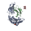 7rzjC  7s79C  7s7dC  7s7eC  7s8aC  7s8eC  7s8fC 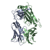 7s8iC 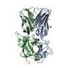 7s8jC S: Starting model for refinement C: citing same article ( |
|---|---|
| Similar structure data |
-Links
 PDBj
PDBj


- Assembly
| Deposited unit | 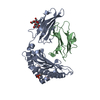
| ||||||||
|---|---|---|---|---|---|---|---|---|---|
| 1 |
| ||||||||
| Unit cell |
|
-Components
| #1: Protein | Mass: 31962.016 Da / Num. of mol.: 1 / Fragment: UNP RESIDUES 25-299 Source method: isolated from a genetically manipulated source Source: (gene. exp.) Homo sapiens (human) / Gene: HLA-B, HLAB / Plasmid: PET30 / Production host:  |
|---|---|
| #2: Protein | Mass: 11879.356 Da / Num. of mol.: 1 / Fragment: UNP residues 21-119 Source method: isolated from a genetically manipulated source Source: (gene. exp.) Homo sapiens (human) / Gene: B2M, CDABP0092, HDCMA22P / Plasmid: PET28a / Production host: |
| #3: Protein/peptide | Mass: 1014.048 Da / Num. of mol.: 1 / Fragment: DOT1L(998-1006) PHOSPHOPEPTIDE / Source method: obtained synthetically / Source: (synth.) Homo sapiens (human)References: UniProt: Q8TEK3, [histone H3]-lysine79 N-trimethyltransferase |
| #4: Polysaccharide | beta-D-fructofuranose-(2-1)-alpha-D-glucopyranose  Source method: isolated from a genetically manipulated source Details: oligosaccharide with reducing-end-to-reducing-end glycosidic bond References: sucrose |
| #5: Water | ChemComp-HOH /  Mass: 18.015 Da / Num. of mol.: 497 / Source method: isolated from a natural source / Formula: H2O Mass: 18.015 Da / Num. of mol.: 497 / Source method: isolated from a natural source / Formula: H2O |
| Has ligand of interest | Y |
| Has protein modification | Y |
-Experimental details
-Experiment
| Experiment | Method: X-RAY DIFFRACTION / Number of used crystals: 1 |
|---|
- Sample preparation
| Crystal | Density Matthews: 2.88 Å3/Da / Density % sol: 57.3 % / Description: rods |
|---|---|
| Crystal grow | Temperature: 290 K / Method: vapor diffusion, sitting drop / pH: 8 Details: 18-24% PEG4000, 0.1 SODIUM CITRATE, 20% ISOPROPANOL, VAPOR DIFFUSION, SITTING DROP, TEMPERATURE |
-Data collection
| Diffraction | Mean temperature: 100 K / Serial crystal experiment: N |
|---|---|
| Diffraction source | Source: SYNCHROTRON / Site: NSLS-II / Beamline: 17-ID-2 / Wavelength: 0.97932 Å |
| Detector | Type: DECTRIS EIGER X 16M / Detector: PIXEL / Date: Mar 4, 2020 / Details: Si(111) MONOCHROMATOR |
| Radiation | Protocol: SINGLE WAVELENGTH / Monochromatic (M) / Laue (L): M / Scattering type: x-ray |
| Radiation wavelength | Wavelength: 0.97932 Å / Relative weight: 1 |
| Reflection | Resolution: 1.88→29.43 Å / Num. obs: 43906 / % possible obs: 99.8 % / Redundancy: 13.2 % / Rmerge(I) obs: 0.112 / Net I/σ(I): 14.7 |
| Reflection shell | Resolution: 1.88→1.92 Å / Redundancy: 13.4 % / Rmerge(I) obs: 0.801 / Mean I/σ(I) obs: 3.5 / Num. unique obs: 3074 / % possible all: 97.2 |
- Processing
| Software |
| ||||||||||||||||||||||||||||||||||||||||||||||||||||||||||||
|---|---|---|---|---|---|---|---|---|---|---|---|---|---|---|---|---|---|---|---|---|---|---|---|---|---|---|---|---|---|---|---|---|---|---|---|---|---|---|---|---|---|---|---|---|---|---|---|---|---|---|---|---|---|---|---|---|---|---|---|---|---|
| Refinement | Method to determine structure: MOLECULAR REPLACEMENT Starting model: 7RZD Resolution: 1.88→29.41 Å / Cor.coef. Fo:Fc: 0.967 / Cor.coef. Fo:Fc free: 0.945 / SU B: 2.988 / SU ML: 0.087 / Cross valid method: THROUGHOUT / σ(F): 0 / ESU R: 0.12 / ESU R Free: 0.124 / Stereochemistry target values: MAXIMUM LIKELIHOOD Details: HYDROGENS HAVE BEEN ADDED IN THE RIDING POSITIONS U VALUES : REFINED INDIVIDUALLY
| ||||||||||||||||||||||||||||||||||||||||||||||||||||||||||||
| Solvent computation | Ion probe radii: 0.8 Å / Shrinkage radii: 0.8 Å / VDW probe radii: 1.3 Å / Solvent model: BABINET MODEL WITH MASK | ||||||||||||||||||||||||||||||||||||||||||||||||||||||||||||
| Displacement parameters | Biso max: 118.95 Å2 / Biso mean: 31.267 Å2 / Biso min: 15.06 Å2
| ||||||||||||||||||||||||||||||||||||||||||||||||||||||||||||
| Refinement step | Cycle: final / Resolution: 1.88→29.41 Å
| ||||||||||||||||||||||||||||||||||||||||||||||||||||||||||||
| Refine LS restraints |
| ||||||||||||||||||||||||||||||||||||||||||||||||||||||||||||
| LS refinement shell | Resolution: 1.88→1.929 Å / Rfactor Rfree error: 0 / Total num. of bins used: 20
|
