 Movie
Movie Controller

[English] 日本語
 Yorodumi
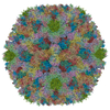
- PDB-7s78: Structure of a cell-entry defective human adenovirus provides ins... -
+ Open data

- Basic information
| Entry | Database: PDB / ID: 7s78 | ||||||
|---|---|---|---|---|---|---|---|
| Title | Structure of a cell-entry defective human adenovirus provides insights into precursor proteins and capsid maturation | ||||||
 Components Components |
| ||||||
 Keywords Keywords | VIRUS / Adenovirus / ts1 mutant / minor protein precursors / virus maturation | ||||||
| Function / homology |  Function and homology information Function and homology informationhexon binding / protein transport along microtubule / viral capsid, decoration / viral procapsid / T=25 icosahedral viral capsid / lysis of host organelle involved in viral entry into host cell / microtubule-dependent intracellular transport of viral material towards nucleus / viral release from host cell / viral life cycle / viral capsid ...hexon binding / protein transport along microtubule / viral capsid, decoration / viral procapsid / T=25 icosahedral viral capsid / lysis of host organelle involved in viral entry into host cell / microtubule-dependent intracellular transport of viral material towards nucleus / viral release from host cell / viral life cycle / viral capsid / host cell / clathrin-dependent endocytosis of virus by host cell / host cell cytoplasm / symbiont entry into host cell / virion attachment to host cell / host cell nucleus / structural molecule activity Similarity search - Function | ||||||
| Biological species |   Human adenovirus C serotype 5 Human adenovirus C serotype 5 | ||||||
| Method | ELECTRON MICROSCOPY / single particle reconstruction / cryo EM / Resolution: 3.72 Å | ||||||
 Authors Authors | Reddy, V.S. / Yu, X. | ||||||
| Funding support |  United States, 1items United States, 1items
| ||||||
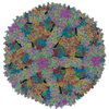
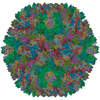
 Citation Citation | Journal: J Mol Biol / Year: 2022 Title: Structure of a Cell Entry Defective Human Adenovirus Provides Insights into Precursor Proteins and Capsid Maturation. Authors: Xiaodi Yu / Tina-Marie Mullen / Vahid Abrishami / Juha T Huiskonen / Glen R Nemerow / Vijay S Reddy /  Abstract: Maturation of adenoviruses is distinguished by proteolytic processing of several interior minor capsid proteins and core proteins by the adenoviral protease and subsequent reorganization of ...Maturation of adenoviruses is distinguished by proteolytic processing of several interior minor capsid proteins and core proteins by the adenoviral protease and subsequent reorganization of adenovirus core. We report the results derived from the icosahedrally averaged cryo-EM structure of a cell entry defective form of adenovirus, designated ts1, at a resolution of 3.7 Å as well as of the localized reconstructions of unique hexons and penton base. The virion structure revealed the structures and organization of precursors of minor capsid proteins, pIIIa, pVI and pVIII, which are closely associated with the hexons on the capsid interior. In addition to a well-ordered helical domain (a.a. 310-397) of pIIIa, highlights of the structure include the precursors of VIII display significantly different structures near the cleavage sites. Moreover, we traced residues 4-96 of the membrane lytic protein (pVI) that includes an amphipathic helix occluded deep in the hexon cavity suggesting the possibility of co-assembly of hexons with the precursors of VI. In addition, we observe a second copy of pVI ordered up to residue L40 in the peripentonal hexons and a few fragments of density corresponding to 2nd and 3rd copies of pVI in other hexons. However, we see no evidence of precursors of VII binding in the hexon cavity. These findings suggest the possibility that differently bound pVI molecules undergo processing at the N-terminal cleavage sites at varying efficiencies, subsequently creating competition between the cleaved and uncleaved forms of VI, followed by reorganization, processing, and release of VI molecules from the hexon cavities. | ||||||
| History |
|
- Structure visualization
| Movie |
Movie viewer |
|---|---|
| Structure viewer | Molecule: MolmilJmol/JSmol |
- Downloads & links
-Download
| PDBx/mmCIF format | 7s78.cif.gz | 2.5 MB | Display | PDBx/mmCIF format |
|---|---|---|---|---|
| PDB format | pdb7s78.ent.gz | Display | PDB format | |
| PDBx/mmJSON format | 7s78.json.gz | Tree view | PDBx/mmJSON format | |
| Others |  Other downloads Other downloads |
-Validation report
| Arichive directory | https://data.pdbj.org/pub/pdb/validation_reports/s7/7s78ftp://data.pdbj.org/pub/pdb/validation_reports/s7/7s78 | HTTPS FTP |
|---|
-Related structure data
| Related structure data |  24881MC M: map data used to model this data C: citing same article ( |
|---|---|
| Similar structure data |

-Links
 PDBj
PDBj


- Assembly
| Deposited unit | 
|
|---|---|
| 1 | x 60
|
| 2 |
|
| 3 | x 5
|
| 4 | x 6
|
| 5 | 
|
| Symmetry | Point symmetry: (Schoenflies symbol: I (icosahedral)) |
-Components
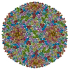
-Protein , 4 types, 26 molecules ABCDEFGHIJKLNPQRSWXYZ01234
| #1: Protein | Mass: 108107.617 Da / Num. of mol.: 12 / Source method: isolated from a natural source / Source: (natural) Human adenovirus C serotype 5 / Strain: Ad2-ts1 / References: UniProt: P04133#2: Protein | | Mass: 63356.602 Da / Num. of mol.: 1 / Source method: isolated from a natural source / Source: (natural) Human adenovirus C serotype 5 / Strain: Ad2-ts1 / References: UniProt: P12538#4: Protein | Mass: 14468.134 Da / Num. of mol.: 4 / Source method: isolated from a natural source / Source: (natural) Human adenovirus C serotype 5 / Strain: Ad2-ts1 / References: UniProt: P03281#6: Protein | Mass: 27027.510 Da / Num. of mol.: 9 / Source method: isolated from a natural source / Source: (natural) Human adenovirus C serotype 5 / Strain: Ad2-ts1 / References: UniProt: P24937 |
|---|
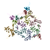
-Pre-hexon-linking protein ... , 2 types, 3 molecules MUV
| #3: Protein | Mass: 65322.805 Da / Num. of mol.: 1 / Source method: isolated from a natural source / Source: (natural) Human adenovirus C serotype 5 / Strain: Ad2-ts1 / References: UniProt: P12537 |
|---|---|
| #5: Protein | Mass: 24710.590 Da / Num. of mol.: 2 / Source method: isolated from a natural source / Source: (natural) Human adenovirus C serotype 5 / Strain: Ad2-ts1 / References: UniProt: P24936 |
-Protein/peptide , 2 types, 2 molecules 56
| #7: Protein/peptide | Mass: 1379.692 Da / Num. of mol.: 1 / Source method: isolated from a natural source / Source: (natural) Human adenovirus C serotype 5 / Strain: Ad2-ts1 |
|---|---|
| #8: Protein/peptide | Mass: 869.063 Da / Num. of mol.: 1 / Source method: isolated from a natural source / Source: (natural) Human adenovirus C serotype 5 / Strain: Ad2-ts1 |
-Experimental details
-Experiment
| Experiment | Method: ELECTRON MICROSCOPY |
|---|---|
| EM experiment | Aggregation state: PARTICLE / 3D reconstruction method: single particle reconstruction |
- Sample preparation
| Component | Name: Human adenovirus C serotype 5 / Type: VIRUS / Entity ID: all / Source: NATURAL |
|---|---|
| Molecular weight | Value: 150 MDa / Experimental value: NO |
| Source (natural) | Organism: Human adenovirus C serotype 5 / Strain: Ad2-ts1 |
| Details of virus | Empty: NO / Enveloped: NO / Isolate: OTHER / Type: VIRION |
| Natural host | Organism: Homo sapiens / Strain: Ad2-ts1 |
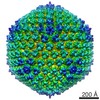
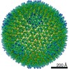
| Virus shell | Name: Capsid / Diameter: 1000 nm / Triangulation number (T number): 25 |
| Buffer solution | pH: 8.1 / Details: 40 mM Tris, pH 8.1 |
| Buffer component | Conc.: 40 mM / Name: Tris |
| Specimen | Conc.: 5 mg/ml / Embedding applied: NO / Shadowing applied: NO / Staining applied: NO / Vitrification applied: YES |
| Specimen support | Details: 20mA / Grid material: COPPER / Grid type: C-flat-1.2/1.3 |
| Vitrification | Cryogen name: ETHANE |
- Electron microscopy imaging
| Experimental equipment |  Model: Titan Krios / Image courtesy: FEI Company |
|---|---|
| Microscopy | Model: FEI TITAN KRIOS |
| Electron gun | Electron source:  FIELD EMISSION GUN / Accelerating voltage: 300 kV / Illumination mode: FLOOD BEAM FIELD EMISSION GUN / Accelerating voltage: 300 kV / Illumination mode: FLOOD BEAM |
| Electron lens | Mode: BRIGHT FIELD / Nominal magnification: 22500 X / Nominal defocus max: 3000 nm / Nominal defocus min: 800 nm / Cs: 2.7 mm / C2 aperture diameter: 100 µm / Alignment procedure: COMA FREE |
| Specimen holder | Cryogen: NITROGEN / Specimen holder model: FEI TITAN KRIOS AUTOGRID HOLDER |
| Image recording | Electron dose: 12 e/Å2 / Detector mode: COUNTING / Film or detector model: GATAN K2 SUMMIT (4k x 4k) / Num. of grids imaged: 2 / Num. of real images: 1510 |
| Image scans | Movie frames/image: 38 |
- Processing
| EM software |
| ||||||||||||||||||||||||||||
|---|---|---|---|---|---|---|---|---|---|---|---|---|---|---|---|---|---|---|---|---|---|---|---|---|---|---|---|---|---|
| CTF correction | Type: PHASE FLIPPING AND AMPLITUDE CORRECTION | ||||||||||||||||||||||||||||
| Particle selection | Num. of particles selected: 21320 | ||||||||||||||||||||||||||||
| Symmetry | Point symmetry: I (icosahedral) | ||||||||||||||||||||||||||||
| 3D reconstruction | Resolution: 3.72 Å / Resolution method: FSC 0.143 CUT-OFF / Num. of particles: 11277 / Algorithm: FOURIER SPACE / Num. of class averages: 18 / Symmetry type: POINT | ||||||||||||||||||||||||||||
| Atomic model building | Protocol: RIGID BODY FIT / Space: REAL | ||||||||||||||||||||||||||||
| Atomic model building | PDB-ID: 3IYN 3iyn Accession code: 3IYN / Source name: PDB / Type: experimental model |
