 Movie
Movie Controller

[English] 日本語
 Yorodumi
- PDB-7s6u: Leishmania infantum Glycogen Synthase Kinase 3 beta bound to AZD5438 -
+ Open data

- Basic information
| Entry | Database: PDB / ID: 7s6u | ||||||||||||
|---|---|---|---|---|---|---|---|---|---|---|---|---|---|
| Title | Leishmania infantum Glycogen Synthase Kinase 3 beta bound to AZD5438 | ||||||||||||
 Components Components | Glycogen synthase kinase 3 | ||||||||||||
 Keywords Keywords | TRANSFERASE/INHIBITOR / Protein kinase / inhibitor / TRANSFERASE / GSK3s / GSK3beta / Leishmania infantum / TRANSFERASE-INHIBITOR complex | ||||||||||||
| Function / homology |  Function and homology information Function and homology informationcell differentiation / non-specific serine/threonine protein kinase / protein serine/threonine kinase activity / signal transduction / ATP binding / nucleus / cytoplasm Similarity search - Function | ||||||||||||
| Biological species |  Leishmania infantum (eukaryote) Leishmania infantum (eukaryote) | ||||||||||||
| Method |  X-RAY DIFFRACTION / SYNCHROTRON / MOLECULAR REPLACEMENT / Resolution: 1.74 Å X-RAY DIFFRACTION / SYNCHROTRON / MOLECULAR REPLACEMENT / Resolution: 1.74 Å | ||||||||||||
 Authors Authors | dos Reis, C.V. / Ramos, P.Z. / Counago, R.M. | ||||||||||||
| Funding support |  Brazil, 3items Brazil, 3items
| ||||||||||||
 Citation Citation | Journal: To Be Published Title: Leishmania infantum Glycogen Synthase Kinase 3 beta bound to AZD5438 Authors: Ramos, P.Z. / dos Reis, C.V. / Counago, R.M. | ||||||||||||
| History |
|
- Structure visualization
| Structure viewer | Molecule: MolmilJmol/JSmol |
|---|
- Downloads & links
-Download
| PDBx/mmCIF format | 7s6u.cif.gz | 154.6 KB | Display | PDBx/mmCIF format |
|---|---|---|---|---|
| PDB format | pdb7s6u.ent.gz | 119.7 KB | Display | PDB format |
| PDBx/mmJSON format | 7s6u.json.gz | Tree view | PDBx/mmJSON format | |
| Others |  Other downloads Other downloads |
-Validation report
| Arichive directory | https://data.pdbj.org/pub/pdb/validation_reports/s6/7s6uftp://data.pdbj.org/pub/pdb/validation_reports/s6/7s6u | HTTPS FTP |
|---|
-Related structure data
| Related structure data | 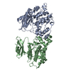 3e3pS S: Starting model for refinement |
|---|---|
| Similar structure data |
-Links
 PDBj
PDBj


- Assembly
| Deposited unit | 
| ||||||||
|---|---|---|---|---|---|---|---|---|---|
| 1 |
| ||||||||
| Unit cell |
|
-Components
| #1: Protein | Mass: 40933.863 Da / Num. of mol.: 1 Source method: isolated from a genetically manipulated source Source: (gene. exp.) Leishmania infantum (eukaryote) / Gene: GSK3, LINF_180007700, LINJ_18_0270 / Production host:  References: UniProt: A4HXQ3, non-specific serine/threonine protein kinase | ||||||||
|---|---|---|---|---|---|---|---|---|---|
| #2: Chemical | ChemComp-FB8 /   Mass: 371.457 Da / Num. of mol.: 1 / Source method: obtained synthetically / Formula: C18H21N5O2S Mass: 371.457 Da / Num. of mol.: 1 / Source method: obtained synthetically / Formula: C18H21N5O2S | ||||||||
| #3: Chemical |   Mass: 62.068 Da / Num. of mol.: 3 / Source method: obtained synthetically / Formula: C2H6O2 Mass: 62.068 Da / Num. of mol.: 3 / Source method: obtained synthetically / Formula: C2H6O2#4: Chemical | ChemComp-CAC / |   Mass: 136.989 Da / Num. of mol.: 1 / Source method: obtained synthetically / Formula: C2H6AsO2 Mass: 136.989 Da / Num. of mol.: 1 / Source method: obtained synthetically / Formula: C2H6AsO2#5: Water | ChemComp-HOH / |  Mass: 18.015 Da / Num. of mol.: 179 / Source method: isolated from a natural source / Formula: H2O Mass: 18.015 Da / Num. of mol.: 179 / Source method: isolated from a natural source / Formula: H2OHas ligand of interest | N | Has protein modification | Y | |
-Experimental details
-Experiment
| Experiment | Method: X-RAY DIFFRACTION / Number of used crystals: 1 |
|---|
- Sample preparation
| Crystal | Density Matthews: 2.32 Å3/Da / Density % sol: 47.05 % |
|---|---|
| Crystal grow | Temperature: 293 K / Method: vapor diffusion, sitting drop / pH: 6 Details: 20% PEG 3350, 0.2 M Sodium bromide, 10% Ethylene glycol, 0.1 M SSB (0.04 M Sodium propionate, 0.02 M Sodium cacodylate trihydrate and 0.04 M Bis-Tris propane) |
-Data collection
| Diffraction | Mean temperature: 100 K / Serial crystal experiment: N |
|---|---|
| Diffraction source | Source: SYNCHROTRON / Site: APS  / Beamline: 24-ID-E / Wavelength: 0.97918 Å / Beamline: 24-ID-E / Wavelength: 0.97918 Å |
| Detector | Type: DECTRIS EIGER X 16M / Detector: PIXEL / Date: Oct 22, 2020 |
| Radiation | Monochromator: Cryogenically-cooled single crystal Si(220) side bounce Protocol: SINGLE WAVELENGTH / Monochromatic (M) / Laue (L): M / Scattering type: x-ray |
| Radiation wavelength | Wavelength: 0.97918 Å / Relative weight: 1 |
| Reflection | Resolution: 1.74→29.26 Å / Num. obs: 38529 / % possible obs: 98.8 % / Redundancy: 4.9 % / Biso Wilson estimate: 26.983 Å2 / CC1/2: 0.998 / Rmerge(I) obs: 0.067 / Rpim(I) all: 0.033 / Rrim(I) all: 0.075 / Net I/σ(I): 11.4 |
| Reflection shell | Resolution: 1.74→1.77 Å / Redundancy: 4.9 % / Rmerge(I) obs: 1.051 / Mean I/σ(I) obs: 1.7 / Num. unique obs: 2106 / CC1/2: 0.514 / Rpim(I) all: 0.51 / Rrim(I) all: 1.173 / % possible all: 98.9 |
- Processing
| Software |
| ||||||||||||||||||||||||||||||||||||||||||||||||||||||||||||
|---|---|---|---|---|---|---|---|---|---|---|---|---|---|---|---|---|---|---|---|---|---|---|---|---|---|---|---|---|---|---|---|---|---|---|---|---|---|---|---|---|---|---|---|---|---|---|---|---|---|---|---|---|---|---|---|---|---|---|---|---|---|
| Refinement | Method to determine structure: MOLECULAR REPLACEMENT Starting model: 3E3P Resolution: 1.74→29.26 Å / Cor.coef. Fo:Fc: 0.97 / Cor.coef. Fo:Fc free: 0.963 / SU B: 5.035 / SU ML: 0.077 / Cross valid method: THROUGHOUT / σ(F): 0 / ESU R: 0.106 / ESU R Free: 0.101 / Stereochemistry target values: MAXIMUM LIKELIHOOD Details: HYDROGENS HAVE BEEN ADDED IN THE RIDING POSITIONS U VALUES : WITH TLS ADDED
| ||||||||||||||||||||||||||||||||||||||||||||||||||||||||||||
| Solvent computation | Ion probe radii: 0.8 Å / Shrinkage radii: 0.8 Å / VDW probe radii: 1.2 Å / Solvent model: MASK | ||||||||||||||||||||||||||||||||||||||||||||||||||||||||||||
| Displacement parameters | Biso max: 88.4 Å2 / Biso mean: 32.377 Å2 / Biso min: 18.79 Å2
| ||||||||||||||||||||||||||||||||||||||||||||||||||||||||||||
| Refinement step | Cycle: final / Resolution: 1.74→29.26 Å
| ||||||||||||||||||||||||||||||||||||||||||||||||||||||||||||
| Refine LS restraints |
| ||||||||||||||||||||||||||||||||||||||||||||||||||||||||||||
| LS refinement shell | Resolution: 1.74→1.785 Å / Rfactor Rfree error: 0 / Total num. of bins used: 20
| ||||||||||||||||||||||||||||||||||||||||||||||||||||||||||||
| Refinement TLS params. | Method: refined / Origin x: 53.8061 Å / Origin y: 22.9359 Å / Origin z: 42.2632 Å
|
