 Movie
Movie Controller

[English] 日本語
 Yorodumi
- PDB-7s3u: Crystal structure of an N-acetyltransferase from Helicobacter pul... -
+ Open data

- Basic information
| Entry | Database: PDB / ID: 7s3u | ||||||
|---|---|---|---|---|---|---|---|
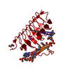
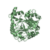
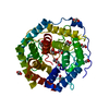
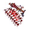
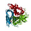
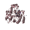
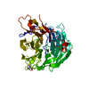
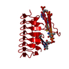
| Title | Crystal structure of an N-acetyltransferase from Helicobacter pullorum in the presence of Coenzyme A and dTDP-3-amino-3,6-dideoxy-D-glucose | ||||||
 Components Components | N-acetyltransferase | ||||||
 Keywords Keywords | TRANSFERASE | ||||||
| Function / homology | COENZYME A / Chem-T3Q Function and homology information Function and homology information | ||||||
| Biological species |  Helicobacter pullorum (bacteria) Helicobacter pullorum (bacteria) | ||||||
| Method |  X-RAY DIFFRACTION / MOLECULAR REPLACEMENT / Resolution: 1.45 Å X-RAY DIFFRACTION / MOLECULAR REPLACEMENT / Resolution: 1.45 Å | ||||||
 Authors Authors | Griffiths, W.A. / Spencer, K.D. / Thoden, J.B. / Holden, H.M. | ||||||
| Funding support |  United States, 1items United States, 1items
| ||||||
 Citation Citation | Journal: Protein Sci. / Year: 2021 Title: Biochemical investigation of an N-acetyltransferase from Helicobacter pullorum. Authors: Griffiths, W.A. / Spencer, K.D. / Thoden, J.B. / Holden, H.M. | ||||||
| History |
|
- Structure visualization
| Structure viewer | Molecule: MolmilJmol/JSmol |
|---|
- Downloads & links
-Download
| PDBx/mmCIF format | 7s3u.cif.gz | 53.1 KB | Display | PDBx/mmCIF format |
|---|---|---|---|---|
| PDB format | pdb7s3u.ent.gz | 34.5 KB | Display | PDB format |
| PDBx/mmJSON format | 7s3u.json.gz | Tree view | PDBx/mmJSON format | |
| Others |  Other downloads Other downloads |
-Validation report
| Arichive directory | https://data.pdbj.org/pub/pdb/validation_reports/s3/7s3uftp://data.pdbj.org/pub/pdb/validation_reports/s3/7s3u | HTTPS FTP |
|---|
-Related structure data
| Related structure data | 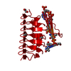 7s3wC  7s41C  7s42C  7s43C 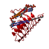 7s44C 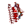 7s45C 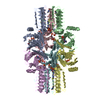 4mzuS S: Starting model for refinement C: citing same article ( |
|---|---|
| Similar structure data |


-Links
 PDBj
PDBj


- Assembly
| Deposited unit | 
| |||||||||
|---|---|---|---|---|---|---|---|---|---|---|
| 1 | 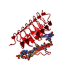
| |||||||||
| Unit cell |
| |||||||||
| Components on special symmetry positions |
|
-Components
| #1: Protein | Mass: 18154.758 Da / Num. of mol.: 1 Source method: isolated from a genetically manipulated source Source: (gene. exp.) Helicobacter pullorum (bacteria) / Strain: NAP8W25 / Gene: BA919_rs02330 / Production host: |
|---|---|
| #2: Chemical | ChemComp-COA /   Mass: 767.534 Da / Num. of mol.: 1 / Source method: obtained synthetically / Formula: C21H36N7O16P3S / Feature type: SUBJECT OF INVESTIGATION Mass: 767.534 Da / Num. of mol.: 1 / Source method: obtained synthetically / Formula: C21H36N7O16P3S / Feature type: SUBJECT OF INVESTIGATION |
| #3: Chemical | ChemComp-T3Q / [(  Mass: 547.345 Da / Num. of mol.: 1 / Source method: obtained synthetically / Formula: C16H27N3O14P2 / Feature type: SUBJECT OF INVESTIGATION Mass: 547.345 Da / Num. of mol.: 1 / Source method: obtained synthetically / Formula: C16H27N3O14P2 / Feature type: SUBJECT OF INVESTIGATION |
| #4: Chemical | ChemComp-EDO /   Mass: 62.068 Da / Num. of mol.: 1 / Source method: obtained synthetically / Formula: C2H6O2 Mass: 62.068 Da / Num. of mol.: 1 / Source method: obtained synthetically / Formula: C2H6O2 |
| #5: Water | ChemComp-HOH /  Mass: 18.015 Da / Num. of mol.: 176 / Source method: isolated from a natural source / Formula: H2O Mass: 18.015 Da / Num. of mol.: 176 / Source method: isolated from a natural source / Formula: H2O |
| Has ligand of interest | Y |
-Experimental details
-Experiment
| Experiment | Method: X-RAY DIFFRACTION / Number of used crystals: 1 |
|---|
- Sample preparation
| Crystal | Density Matthews: 2.51 Å3/Da / Density % sol: 50.91 % |
|---|---|
| Crystal grow | Temperature: 293 K / Method: vapor diffusion, hanging drop / pH: 8 Details: 16-20% PEG-3350, 200 mM KCl, 5 mM dTDP-3-amino-3,6-dideoxy-D-glucose, 5 mM Coenzyme A, 100 mM HEPPS |
-Data collection
| Diffraction | Mean temperature: 100 K / Serial crystal experiment: N |
|---|---|
| Diffraction source | Source: SEALED TUBE / Type: BRUKER D8 QUEST / Wavelength: 1.5418 Å |
| Detector | Type: Bruker PHOTON II / Detector: PIXEL / Date: Aug 27, 2020 |
| Radiation | Protocol: SINGLE WAVELENGTH / Monochromatic (M) / Laue (L): M / Scattering type: x-ray |
| Radiation wavelength | Wavelength: 1.5418 Å / Relative weight: 1 |
| Reflection | Resolution: 1.45→50 Å / Num. obs: 32307 / % possible obs: 99.8 % / Observed criterion σ(F): 0 / Observed criterion σ(I): 0 / Redundancy: 14.3 % / Rsym value: 0.044 / Net I/σ(I): 18.7 |
| Reflection shell | Resolution: 1.456→1.55 Å / Redundancy: 8.8 % / Mean I/σ(I) obs: 5.1 / Num. unique obs: 5844 / Rsym value: 0.214 / % possible all: 99 |
- Processing
| Software |
| ||||||||||||||||||||||||||||||||||||||||||||||||||||||||||||
|---|---|---|---|---|---|---|---|---|---|---|---|---|---|---|---|---|---|---|---|---|---|---|---|---|---|---|---|---|---|---|---|---|---|---|---|---|---|---|---|---|---|---|---|---|---|---|---|---|---|---|---|---|---|---|---|---|---|---|---|---|---|
| Refinement | Method to determine structure: MOLECULAR REPLACEMENT Starting model: 4mzu Resolution: 1.45→36.43 Å / Cor.coef. Fo:Fc: 0.962 / Cor.coef. Fo:Fc free: 0.957 / SU B: 1.155 / SU ML: 0.042 / Cross valid method: THROUGHOUT / σ(F): 0 / ESU R: 0.06 / ESU R Free: 0.06 / Stereochemistry target values: MAXIMUM LIKELIHOOD Details: HYDROGENS HAVE BEEN ADDED IN THE RIDING POSITIONS U VALUES : REFINED INDIVIDUALLY
| ||||||||||||||||||||||||||||||||||||||||||||||||||||||||||||
| Solvent computation | Ion probe radii: 0.8 Å / Shrinkage radii: 0.8 Å / VDW probe radii: 1.2 Å / Solvent model: MASK | ||||||||||||||||||||||||||||||||||||||||||||||||||||||||||||
| Displacement parameters | Biso max: 72.85 Å2 / Biso mean: 11.482 Å2 / Biso min: 4.05 Å2
| ||||||||||||||||||||||||||||||||||||||||||||||||||||||||||||
| Refinement step | Cycle: final / Resolution: 1.45→36.43 Å
| ||||||||||||||||||||||||||||||||||||||||||||||||||||||||||||
| Refine LS restraints |
| ||||||||||||||||||||||||||||||||||||||||||||||||||||||||||||
| LS refinement shell | Resolution: 1.45→1.486 Å / Rfactor Rfree error: 0
|
