 Movie
Movie Controller

[English] 日本語
 Yorodumi
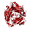
- PDB-7rwh: Crystal structure of human methionine adenosyltransferase 2A (MAT... -
+ Open data

- Basic information
| Entry | Database: PDB / ID: 7rwh | ||||||
|---|---|---|---|---|---|---|---|
| Title | Crystal structure of human methionine adenosyltransferase 2A (MAT2A) in complex with SAM and allosteric inhibitor AGI-41998 | ||||||
 Components Components | S-adenosylmethionine synthase isoform type-2 | ||||||
 Keywords Keywords | TRANSFERASE/INHIBITOR / METHIONINE ADENOSYLTRANSFERASE / SAM / ALLOSTERIC INHIBITOR / TRANSFERASE / TRANSFERASE-INHIBITOR COMPLEX | ||||||
| Function / homology |  Function and homology information Function and homology informationmethionine adenosyltransferase complex / methionine adenosyltransferase / methionine adenosyltransferase activity / S-adenosylmethionine biosynthetic process / protein heterooligomerization / Methylation / cellular response to methionine / protein hexamerization / small molecule binding / one-carbon metabolic process ...methionine adenosyltransferase complex / methionine adenosyltransferase / methionine adenosyltransferase activity / S-adenosylmethionine biosynthetic process / protein heterooligomerization / Methylation / cellular response to methionine / protein hexamerization / small molecule binding / one-carbon metabolic process / positive regulation of TORC1 signaling / cellular response to leukemia inhibitory factor / ATP binding / metal ion binding / identical protein binding / cytosol Similarity search - Function | ||||||
| Biological species |  Homo sapiens (human) Homo sapiens (human) | ||||||
| Method |  X-RAY DIFFRACTION / SYNCHROTRON / MOLECULAR REPLACEMENT / Resolution: 1.17 Å X-RAY DIFFRACTION / SYNCHROTRON / MOLECULAR REPLACEMENT / Resolution: 1.17 Å | ||||||
 Authors Authors | Jin, L. / Padyana, A.K. | ||||||
| Funding support | 1items
| ||||||
 Citation Citation | Journal: J.Med.Chem. / Year: 2022 Title: Leveraging Structure-Based Drug Design to Identify Next-Generation MAT2A Inhibitors, Including Brain-Penetrant and Peripherally Efficacious Leads. Authors: Li, M. / Konteatis, Z. / Nagaraja, N. / Chen, Y. / Zhou, S. / Ma, G. / Gross, S. / Marjon, K. / Hyer, M.L. / Mandley, E. / Lein, M. / Padyana, A.K. / Jin, L. / Tong, S. / Peters, R. / ...Authors: Li, M. / Konteatis, Z. / Nagaraja, N. / Chen, Y. / Zhou, S. / Ma, G. / Gross, S. / Marjon, K. / Hyer, M.L. / Mandley, E. / Lein, M. / Padyana, A.K. / Jin, L. / Tong, S. / Peters, R. / Murtie, J. / Travins, J. / Medeiros, M. / Liu, P. / Frank, V. / Judd, E.T. / Biller, S.A. / Marks, K.M. / Sui, Z. / Reznik, S.K. #1: Journal: J.MED.CHEM. / Year: 2021Title: DISCOVERY OF AG-270, A FIRST-IN-CLASS ORAL MAT2A INHIBITOR DISCOVERY OF AG-270, A FIRST-IN-CLASS ORAL MAT2A INHIBITOR Authors: Konteatis, Z. / Travins, J. | ||||||
| History |
|
- Structure visualization
| Structure viewer | Molecule: MolmilJmol/JSmol |
|---|
- Downloads & links
-Download
| PDBx/mmCIF format | 7rwh.cif.gz | 325.6 KB | Display | PDBx/mmCIF format |
|---|---|---|---|---|
| PDB format | pdb7rwh.ent.gz | 217.2 KB | Display | PDB format |
| PDBx/mmJSON format | 7rwh.json.gz | Tree view | PDBx/mmJSON format | |
| Others |  Other downloads Other downloads |
-Validation report
| Arichive directory | https://data.pdbj.org/pub/pdb/validation_reports/rw/7rwhftp://data.pdbj.org/pub/pdb/validation_reports/rw/7rwh | HTTPS FTP |
|---|
-Related structure data
| Related structure data | 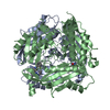 7rw5C 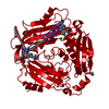 7rw7C 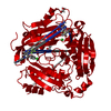 7rwgC 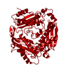 2p02S C: citing same article ( S: Starting model for refinement |
|---|---|
| Similar structure data |
-Links
 PDBj
PDBj
- Assembly
| Deposited unit | 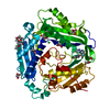
| ||||||||||||||||||||||||
|---|---|---|---|---|---|---|---|---|---|---|---|---|---|---|---|---|---|---|---|---|---|---|---|---|---|
| 1 | 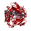
| ||||||||||||||||||||||||
| Unit cell |
| ||||||||||||||||||||||||
| Components on special symmetry positions |
|
-Components
-Protein , 1 types, 1 molecules A
| #1: Protein | Mass: 43807.703 Da / Num. of mol.: 1 Source method: isolated from a genetically manipulated source Source: (gene. exp.) Homo sapiens (human) / Gene: MAT2A, AMS2, MATA2 / Production host:  |
|---|
-Non-polymers , 8 types, 542 molecules 


| #2: Chemical | ChemComp-SAM /  Mass: 398.437 Da / Num. of mol.: 1 / Source method: obtained synthetically / Formula: C15H22N6O5S Mass: 398.437 Da / Num. of mol.: 1 / Source method: obtained synthetically / Formula: C15H22N6O5S | ||||||||||
|---|---|---|---|---|---|---|---|---|---|---|---|
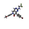
| #3: Chemical | ChemComp-7UI /  Mass: 505.287 Da / Num. of mol.: 1 / Source method: obtained synthetically / Formula: C22H16BrF3N4O2 / Feature type: SUBJECT OF INVESTIGATION Mass: 505.287 Da / Num. of mol.: 1 / Source method: obtained synthetically / Formula: C22H16BrF3N4O2 / Feature type: SUBJECT OF INVESTIGATION | ||||||||||
| #4: Chemical | ChemComp-EDO /  Mass: 62.068 Da / Num. of mol.: 4 / Source method: obtained synthetically / Formula: C2H6O2 Mass: 62.068 Da / Num. of mol.: 4 / Source method: obtained synthetically / Formula: C2H6O2#5: Chemical | ChemComp-GOL / |  Mass: 92.094 Da / Num. of mol.: 1 / Source method: obtained synthetically / Formula: C3H8O3 Mass: 92.094 Da / Num. of mol.: 1 / Source method: obtained synthetically / Formula: C3H8O3#6: Chemical | ChemComp-1PE / |  Mass: 238.278 Da / Num. of mol.: 1 / Source method: obtained synthetically / Formula: C10H22O6 / Comment: precipitant*YM Mass: 238.278 Da / Num. of mol.: 1 / Source method: obtained synthetically / Formula: C10H22O6 / Comment: precipitant*YM#7: Chemical | ChemComp-CL / |  Mass: 35.453 Da / Num. of mol.: 1 / Source method: obtained synthetically / Formula: Cl Mass: 35.453 Da / Num. of mol.: 1 / Source method: obtained synthetically / Formula: Cl#8: Chemical |  Mass: 24.305 Da / Num. of mol.: 3 / Source method: obtained synthetically / Formula: Mg Mass: 24.305 Da / Num. of mol.: 3 / Source method: obtained synthetically / Formula: Mg#9: Water | ChemComp-HOH / | Mass: 18.015 Da / Num. of mol.: 530 / Source method: isolated from a natural source / Formula: H2O |
-Details
| Has ligand of interest | Y |
|---|
-Experimental details
-Experiment
| Experiment | Method: X-RAY DIFFRACTION / Number of used crystals: 1 |
|---|
- Sample preparation
| Crystal | Density Matthews: 2.15 Å3/Da / Density % sol: 42.75 % |
|---|---|
| Crystal grow | Temperature: 291 K / Method: vapor diffusion, hanging drop / pH: 8 Details: 0.2 M LiCl, 0.1 M Tris pH 8.0, 18%-20% PEG6000, 10% Ethylene Glycol |
-Data collection
| Diffraction | Mean temperature: 100 K / Serial crystal experiment: N |
|---|---|
| Diffraction source | Source: SYNCHROTRON / Site: APS  / Beamline: 21-ID-D / Wavelength: 1.1271 Å / Beamline: 21-ID-D / Wavelength: 1.1271 Å |
| Detector | Type: DECTRIS EIGER X 9M / Detector: PIXEL / Date: Jul 13, 2019 |
| Radiation | Protocol: SINGLE WAVELENGTH / Monochromatic (M) / Laue (L): M / Scattering type: x-ray |
| Radiation wavelength | Wavelength: 1.1271 Å / Relative weight: 1 |
| Reflection | Resolution: 1.17→36.63 Å / Num. obs: 116890 / % possible obs: 92.3 % / Redundancy: 11.6 % / Biso Wilson estimate: 9.25 Å2 / Rmerge(I) obs: 0.114 / Net I/σ(I): 12.1 |
| Reflection shell | Resolution: 1.17→1.2 Å / Rmerge(I) obs: 0.675 / Mean I/σ(I) obs: 2.2 / Num. unique obs: 3998 |
- Processing
| Software |
| |||||||||||||||||||||||||||||||||||||||||||||||||||||||||||||||||||||||||||||||||||||||||||||||||||||||||||||||||||||||||||||||||||||||||||||||||||||||||||||||||||||||||||||||||||||||||||||||||||||||||||||||||||||||||
|---|---|---|---|---|---|---|---|---|---|---|---|---|---|---|---|---|---|---|---|---|---|---|---|---|---|---|---|---|---|---|---|---|---|---|---|---|---|---|---|---|---|---|---|---|---|---|---|---|---|---|---|---|---|---|---|---|---|---|---|---|---|---|---|---|---|---|---|---|---|---|---|---|---|---|---|---|---|---|---|---|---|---|---|---|---|---|---|---|---|---|---|---|---|---|---|---|---|---|---|---|---|---|---|---|---|---|---|---|---|---|---|---|---|---|---|---|---|---|---|---|---|---|---|---|---|---|---|---|---|---|---|---|---|---|---|---|---|---|---|---|---|---|---|---|---|---|---|---|---|---|---|---|---|---|---|---|---|---|---|---|---|---|---|---|---|---|---|---|---|---|---|---|---|---|---|---|---|---|---|---|---|---|---|---|---|---|---|---|---|---|---|---|---|---|---|---|---|---|---|---|---|---|---|---|---|---|---|---|---|---|---|---|---|---|---|---|---|---|
| Refinement | Method to determine structure: MOLECULAR REPLACEMENT Starting model: 2P02 Resolution: 1.17→35.95 Å / SU ML: 0.0743 / Cross valid method: FREE R-VALUE / σ(F): 1.35 / Phase error: 13.1522 Stereochemistry target values: GeoStd + Monomer Library + CDL v1.2
| |||||||||||||||||||||||||||||||||||||||||||||||||||||||||||||||||||||||||||||||||||||||||||||||||||||||||||||||||||||||||||||||||||||||||||||||||||||||||||||||||||||||||||||||||||||||||||||||||||||||||||||||||||||||||
| Solvent computation | Shrinkage radii: 0.9 Å / VDW probe radii: 1.11 Å / Solvent model: FLAT BULK SOLVENT MODEL | |||||||||||||||||||||||||||||||||||||||||||||||||||||||||||||||||||||||||||||||||||||||||||||||||||||||||||||||||||||||||||||||||||||||||||||||||||||||||||||||||||||||||||||||||||||||||||||||||||||||||||||||||||||||||
| Displacement parameters | Biso mean: 14.78 Å2 | |||||||||||||||||||||||||||||||||||||||||||||||||||||||||||||||||||||||||||||||||||||||||||||||||||||||||||||||||||||||||||||||||||||||||||||||||||||||||||||||||||||||||||||||||||||||||||||||||||||||||||||||||||||||||
| Refinement step | Cycle: LAST / Resolution: 1.17→35.95 Å
| |||||||||||||||||||||||||||||||||||||||||||||||||||||||||||||||||||||||||||||||||||||||||||||||||||||||||||||||||||||||||||||||||||||||||||||||||||||||||||||||||||||||||||||||||||||||||||||||||||||||||||||||||||||||||
| Refine LS restraints |
| |||||||||||||||||||||||||||||||||||||||||||||||||||||||||||||||||||||||||||||||||||||||||||||||||||||||||||||||||||||||||||||||||||||||||||||||||||||||||||||||||||||||||||||||||||||||||||||||||||||||||||||||||||||||||
| LS refinement shell |
| |||||||||||||||||||||||||||||||||||||||||||||||||||||||||||||||||||||||||||||||||||||||||||||||||||||||||||||||||||||||||||||||||||||||||||||||||||||||||||||||||||||||||||||||||||||||||||||||||||||||||||||||||||||||||
| Refinement TLS params. | Method: refined / Refine-ID: X-RAY DIFFRACTION
| |||||||||||||||||||||||||||||||||||||||||||||||||||||||||||||||||||||||||||||||||||||||||||||||||||||||||||||||||||||||||||||||||||||||||||||||||||||||||||||||||||||||||||||||||||||||||||||||||||||||||||||||||||||||||
| Refinement TLS group |
|
