 Movie
Movie Controller

[English] 日本語
 Yorodumi
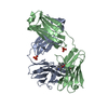
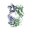
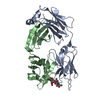
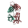
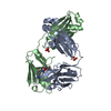
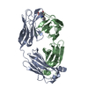
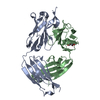
- PDB-7rtp: Structure of full-length human lambda-6A light chain JTO in compl... -
+ Open data

- Basic information
| Entry | Database: PDB / ID: 7rtp | ||||||
|---|---|---|---|---|---|---|---|
| Title | Structure of full-length human lambda-6A light chain JTO in complex with urea stabilizer 20 [1-(2-(7-(diethylamino)-4-methyl-2-oxo-2H-chromen-3-yl)ethyl)-3-(pyridin-3-ylmethyl)urea] | ||||||
 Components Components | JTO light chain | ||||||
 Keywords Keywords | IMMUNE SYSTEM / amyloidosis | ||||||
| Function / homology | Chem-NY9 / PHOSPHATE ION Function and homology information Function and homology information | ||||||
| Biological species |  Homo sapiens (human) Homo sapiens (human) | ||||||
| Method |  X-RAY DIFFRACTION / SYNCHROTRON / MOLECULAR REPLACEMENT / Resolution: 2.09 Å X-RAY DIFFRACTION / SYNCHROTRON / MOLECULAR REPLACEMENT / Resolution: 2.09 Å | ||||||
 Authors Authors | Yan, N.L. / Wilson, I.A. / Kelly, J.W. | ||||||
| Funding support |  United States, 1items United States, 1items
| ||||||
 Citation Citation | Journal: Bioorg.Med.Chem.Lett. / Year: 2022 Title: Amyloidogenic immunoglobulin light chain kinetic stabilizers comprising a simple urea linker module reveal a novel binding sub-site. Authors: Yan, N.L. / Nair, R. / Chu, A. / Wilson, I.A. / Johnson, K.A. / Morgan, G.J. / Kelly, J.W. | ||||||
| History |
|
- Structure visualization
| Structure viewer | Molecule: MolmilJmol/JSmol |
|---|
- Downloads & links
-Download
| PDBx/mmCIF format | 7rtp.cif.gz | 106 KB | Display | PDBx/mmCIF format |
|---|---|---|---|---|
| PDB format | pdb7rtp.ent.gz | 78.7 KB | Display | PDB format |
| PDBx/mmJSON format | 7rtp.json.gz | Tree view | PDBx/mmJSON format | |
| Others |  Other downloads Other downloads |
-Validation report
| Summary document | 7rtp_validation.pdf.gz | 686 KB | Display | wwPDB validaton report |
|---|---|---|---|---|
| Full document | 7rtp_full_validation.pdf.gz | 688.4 KB | Display | |
| Data in XML | 7rtp_validation.xml.gz | 20.7 KB | Display | |
| Data in CIF | 7rtp_validation.cif.gz | 30.8 KB | Display | |
| Arichive directory | https://data.pdbj.org/pub/pdb/validation_reports/rt/7rtpftp://data.pdbj.org/pub/pdb/validation_reports/rt/7rtp | HTTPS FTP |
-Related structure data
| Related structure data | 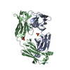 6mg5S S: Starting model for refinement |
|---|---|
| Similar structure data |


-Links
 PDBj
PDBj

- Assembly
| Deposited unit | 
| ||||||||
|---|---|---|---|---|---|---|---|---|---|
| 1 |
| ||||||||
| Unit cell |
|
-Components
| #1: Antibody | Mass: 23211.543 Da / Num. of mol.: 2 Source method: isolated from a genetically manipulated source Source: (gene. exp.) Homo sapiens (human) / Production host:  #2: Chemical |   Mass: 94.971 Da / Num. of mol.: 2 / Source method: obtained synthetically / Formula: PO4 Mass: 94.971 Da / Num. of mol.: 2 / Source method: obtained synthetically / Formula: PO4#3: Chemical | ChemComp-NY9 / | 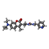  Mass: 408.493 Da / Num. of mol.: 1 / Source method: obtained synthetically / Formula: C23H28N4O3 / Feature type: SUBJECT OF INVESTIGATION Mass: 408.493 Da / Num. of mol.: 1 / Source method: obtained synthetically / Formula: C23H28N4O3 / Feature type: SUBJECT OF INVESTIGATION#4: Water | ChemComp-HOH / |  Mass: 18.015 Da / Num. of mol.: 357 / Source method: isolated from a natural source / Formula: H2O Mass: 18.015 Da / Num. of mol.: 357 / Source method: isolated from a natural source / Formula: H2OHas ligand of interest | Y | |
|---|
-Experimental details
-Experiment
| Experiment | Method: X-RAY DIFFRACTION / Number of used crystals: 1 |
|---|
- Sample preparation
| Crystal | Density Matthews: 2.61 Å3/Da / Density % sol: 52.95 % |
|---|---|
| Crystal grow | Temperature: 296 K / Method: vapor diffusion, sitting drop / Details: 20% PEG 3350 and 0.25 M NH4H2PO4 at 23 degrees C |
-Data collection
| Diffraction | Mean temperature: 100 K / Serial crystal experiment: N |
|---|---|
| Diffraction source | Source: SYNCHROTRON / Site: APS / Beamline: 23-ID-B / Wavelength: 1.0332 Å |
| Detector | Type: DECTRIS EIGER X 16M / Detector: PIXEL / Date: Jun 8, 2019 |
| Radiation | Protocol: SINGLE WAVELENGTH / Monochromatic (M) / Laue (L): M / Scattering type: x-ray |
| Radiation wavelength | Wavelength: 1.0332 Å / Relative weight: 1 |
| Reflection | Resolution: 2.09→47.37 Å / Num. obs: 29655 / % possible obs: 99.8 % / Redundancy: 12.9 % / CC1/2: 1 / Rpim(I) all: 0.034 / Rsym value: 0.12 / Net I/σ(I): 14.9 |
| Reflection shell | Resolution: 2.09→2.2 Å / Mean I/σ(I) obs: 3.7 / Num. unique obs: 4230 / CC1/2: 0.92 / Rpim(I) all: 0.21 / Rsym value: 0.72 |
- Processing
| Software |
| ||||||||||||||||||||||||||||||||||||||||||||||||||||||||||||
|---|---|---|---|---|---|---|---|---|---|---|---|---|---|---|---|---|---|---|---|---|---|---|---|---|---|---|---|---|---|---|---|---|---|---|---|---|---|---|---|---|---|---|---|---|---|---|---|---|---|---|---|---|---|---|---|---|---|---|---|---|---|
| Refinement | Method to determine structure: MOLECULAR REPLACEMENT Starting model: 6MG5 Resolution: 2.09→47.37 Å / Cor.coef. Fo:Fc: 0.959 / Cor.coef. Fo:Fc free: 0.921 / Cross valid method: THROUGHOUT / σ(F): 0 / ESU R: 0.199 / ESU R Free: 0.183 / Stereochemistry target values: MAXIMUM LIKELIHOOD Details: HYDROGENS HAVE BEEN ADDED IN THE RIDING POSITIONS U VALUES : REFINED INDIVIDUALLY
| ||||||||||||||||||||||||||||||||||||||||||||||||||||||||||||
| Solvent computation | Ion probe radii: 0.8 Å / Shrinkage radii: 0.8 Å / VDW probe radii: 1.2 Å / Solvent model: MASK | ||||||||||||||||||||||||||||||||||||||||||||||||||||||||||||
| Displacement parameters | Biso max: 156.13 Å2 / Biso mean: 30.592 Å2 / Biso min: 18.17 Å2
| ||||||||||||||||||||||||||||||||||||||||||||||||||||||||||||
| Refinement step | Cycle: final / Resolution: 2.09→47.37 Å
| ||||||||||||||||||||||||||||||||||||||||||||||||||||||||||||
| Refine LS restraints |
| ||||||||||||||||||||||||||||||||||||||||||||||||||||||||||||
| LS refinement shell | Resolution: 2.09→2.141 Å / Rfactor Rfree error: 0
|
