 Movie
Movie Controller


+ Open data

- Basic information
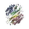
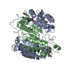
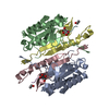
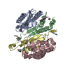
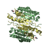
| Entry | Database: PDB / ID: 7rnb | ||||||||||||
|---|---|---|---|---|---|---|---|---|---|---|---|---|---|
| Title | Crystal structure of caspase-3 with inhibitor Ac-VDRVD-CHO | ||||||||||||
 Components Components |
| ||||||||||||
 Keywords Keywords | HYDROLASE / Hydrolase/Hydrolase Inhibitor | ||||||||||||
| Function / homology |  Function and homology information Function and homology informationcaspase-3 / phospholipase A2 activator activity / Stimulation of the cell death response by PAK-2p34 / anterior neural tube closure / intrinsic apoptotic signaling pathway in response to osmotic stress / leukocyte apoptotic process / positive regulation of pyroptotic inflammatory response / glial cell apoptotic process / NADE modulates death signalling / luteolysis ...caspase-3 / phospholipase A2 activator activity / Stimulation of the cell death response by PAK-2p34 / anterior neural tube closure / intrinsic apoptotic signaling pathway in response to osmotic stress / leukocyte apoptotic process / positive regulation of pyroptotic inflammatory response / glial cell apoptotic process / NADE modulates death signalling / luteolysis / response to cobalt ion / cellular response to staurosporine / cyclin-dependent protein serine/threonine kinase inhibitor activity / death-inducing signaling complex / Apoptosis induced DNA fragmentation / Apoptotic cleavage of cell adhesion proteins / Caspase activation via Dependence Receptors in the absence of ligand / SMAC, XIAP-regulated apoptotic response / Activation of caspases through apoptosome-mediated cleavage / Signaling by Hippo / SMAC (DIABLO) binds to IAPs / SMAC(DIABLO)-mediated dissociation of IAP:caspase complexes / axonal fasciculation / regulation of synaptic vesicle cycle / death receptor binding / fibroblast apoptotic process / epithelial cell apoptotic process / platelet formation / Other interleukin signaling / response to anesthetic / execution phase of apoptosis / negative regulation of cytokine production / positive regulation of amyloid-beta formation / Apoptotic cleavage of cellular proteins / negative regulation of B cell proliferation / pyroptotic inflammatory response / neurotrophin TRK receptor signaling pathway / negative regulation of activated T cell proliferation / negative regulation of cell cycle / response to tumor necrosis factor / T cell homeostasis / B cell homeostasis / Pyroptosis / cell fate commitment / regulation of macroautophagy / Caspase-mediated cleavage of cytoskeletal proteins / response to X-ray / response to amino acid / response to glucose / response to UV / keratinocyte differentiation / Degradation of the extracellular matrix / striated muscle cell differentiation / intrinsic apoptotic signaling pathway / response to glucocorticoid / protein maturation / erythrocyte differentiation / response to nicotine / hippocampus development / apoptotic signaling pathway / enzyme activator activity / protein catabolic process / response to hydrogen peroxide / sensory perception of sound / regulation of protein stability / protein processing / response to wounding / neuron differentiation / response to estradiol / peptidase activity / positive regulation of neuron apoptotic process / heart development / protease binding / neuron apoptotic process / response to lipopolysaccharide / aspartic-type endopeptidase activity / learning or memory / response to hypoxia / postsynaptic density / response to xenobiotic stimulus / cysteine-type endopeptidase activity / neuronal cell body / apoptotic process / DNA damage response / protein-containing complex binding / glutamatergic synapse / proteolysis / nucleoplasm / nucleus / cytoplasm / cytosol Similarity search - Function | ||||||||||||
| Biological species |  Homo sapiens (human) Homo sapiens (human)synthetic construct (others) | ||||||||||||
| Method |  X-RAY DIFFRACTION / SYNCHROTRON / MOLECULAR REPLACEMENT / Resolution: 1.75 Å X-RAY DIFFRACTION / SYNCHROTRON / MOLECULAR REPLACEMENT / Resolution: 1.75 Å | ||||||||||||
 Authors Authors | McCue, W. / Finzel, B.C. | ||||||||||||
| Funding support |  United States, 1items United States, 1items
| ||||||||||||
 Citation Citation | Journal: Acs Pharmacol Transl Sci / Year: 2022 Title: Structure-Based Design and Biological Evaluation of Novel Caspase-2 Inhibitors Based on the Peptide AcVDVAD-CHO and the Caspase-2-Mediated Tau Cleavage Sequence YKPVD314. Authors: Bresinsky, M. / Strasser, J.M. / Vallaster, B. / Liu, P. / McCue, W.M. / Fuller, J. / Hubmann, A. / Singh, G. / Nelson, K.M. / Cuellar, M.E. / Wilmot, C.M. / Finzel, B.C. / Ashe, K.H. / ...Authors: Bresinsky, M. / Strasser, J.M. / Vallaster, B. / Liu, P. / McCue, W.M. / Fuller, J. / Hubmann, A. / Singh, G. / Nelson, K.M. / Cuellar, M.E. / Wilmot, C.M. / Finzel, B.C. / Ashe, K.H. / Walters, M.A. / Pockes, S. | ||||||||||||
| History |
|
- Structure visualization
| Structure viewer | Molecule: MolmilJmol/JSmol |
|---|
- Downloads & links
-Download
| PDBx/mmCIF format | 7rnb.cif.gz | 141.3 KB | Display | PDBx/mmCIF format |
|---|---|---|---|---|
| PDB format | pdb7rnb.ent.gz | 88.3 KB | Display | PDB format |
| PDBx/mmJSON format | 7rnb.json.gz | Tree view | PDBx/mmJSON format | |
| Others |  Other downloads Other downloads |
-Validation report
| Summary document | 7rnb_validation.pdf.gz | 447.1 KB | Display | wwPDB validaton report |
|---|---|---|---|---|
| Full document | 7rnb_full_validation.pdf.gz | 448.1 KB | Display | |
| Data in XML | 7rnb_validation.xml.gz | 21 KB | Display | |
| Data in CIF | 7rnb_validation.cif.gz | 30.5 KB | Display | |
| Arichive directory | https://data.pdbj.org/pub/pdb/validation_reports/rn/7rnbftp://data.pdbj.org/pub/pdb/validation_reports/rn/7rnb | HTTPS FTP |
-Related structure data
| Related structure data |  7rn7C  7rn8C 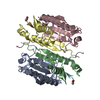 7rn9C  7rndC  7rneC  7rnfC  7seoC  2h65S S: Starting model for refinement C: citing same article ( |
|---|---|
| Similar structure data |


-Links
 PDBj
PDBj


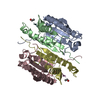
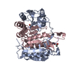
- Assembly
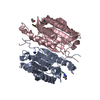
| Deposited unit | 
| ||||||||||||
|---|---|---|---|---|---|---|---|---|---|---|---|---|---|
| 1 |
| ||||||||||||
| Unit cell |
|
-Components
| #1: Protein | Mass: 16067.288 Da / Num. of mol.: 2 Source method: isolated from a genetically manipulated source Source: (gene. exp.) Homo sapiens (human) / Gene: CASP3, CPP32 / Production host:  #2: Protein | Mass: 11257.953 Da / Num. of mol.: 2 Source method: isolated from a genetically manipulated source Source: (gene. exp.) Homo sapiens (human) / Gene: CASP3, CPP32 / Production host: #3: Protein/peptide | Mass: 613.684 Da / Num. of mol.: 2 / Source method: obtained synthetically / Source: (synth.) synthetic construct (others) #4: Water | ChemComp-HOH / |  Mass: 18.015 Da / Num. of mol.: 260 / Source method: isolated from a natural source / Formula: H2O Mass: 18.015 Da / Num. of mol.: 260 / Source method: isolated from a natural source / Formula: H2OHas ligand of interest | Y | Has protein modification | Y | |
|---|
-Experimental details
-Experiment
| Experiment | Method: X-RAY DIFFRACTION / Number of used crystals: 1 |
|---|
- Sample preparation
| Crystal | Density Matthews: 2.59 Å3/Da / Density % sol: 52.49 % |
|---|---|
| Crystal grow | Temperature: 293 K / Method: vapor diffusion, hanging drop Details: 15% PEG 6000, 5% glycerol (v:v), 100 mM sodium citrate pH 5.3, 10 mM DTT and 3 mM NaN3 |
-Data collection
| Diffraction | Mean temperature: 100 K / Serial crystal experiment: N |
|---|---|
| Diffraction source | Source: SYNCHROTRON / Site: APS / Beamline: 17-ID / Wavelength: 1 Å |
| Detector | Type: DECTRIS EIGER2 X 9M / Detector: PIXEL / Date: Mar 15, 2020 |
| Radiation | Protocol: SINGLE WAVELENGTH / Monochromatic (M) / Laue (L): M / Scattering type: x-ray |
| Radiation wavelength | Wavelength: 1 Å / Relative weight: 1 |
| Reflection | Resolution: 1.75→64.91 Å / Num. obs: 56330 / % possible obs: 95.9 % / Redundancy: 10.6 % / CC1/2: 0.999 / Rmerge(I) obs: 0.092 / Rpim(I) all: 0.03 / Rrim(I) all: 0.097 / Net I/σ(I): 16.9 / Num. measured all: 596208 / Scaling rejects: 126 |
| Reflection shell | Resolution: 1.75→1.84 Å / Redundancy: 10.3 % / Rmerge(I) obs: 0.729 / Num. unique obs: 8435 / CC1/2: 0.924 / Rpim(I) all: 0.235 / Rrim(I) all: 0.767 / % possible all: 98.9 |
- Processing
| Software |
| |||||||||||||||||||||||||||||||||||||||||||||||||||||||||||||||||||||||||||||||||||||||||||||||||||||||||||||||||||||||||||||||||||||||||||||||||||
|---|---|---|---|---|---|---|---|---|---|---|---|---|---|---|---|---|---|---|---|---|---|---|---|---|---|---|---|---|---|---|---|---|---|---|---|---|---|---|---|---|---|---|---|---|---|---|---|---|---|---|---|---|---|---|---|---|---|---|---|---|---|---|---|---|---|---|---|---|---|---|---|---|---|---|---|---|---|---|---|---|---|---|---|---|---|---|---|---|---|---|---|---|---|---|---|---|---|---|---|---|---|---|---|---|---|---|---|---|---|---|---|---|---|---|---|---|---|---|---|---|---|---|---|---|---|---|---|---|---|---|---|---|---|---|---|---|---|---|---|---|---|---|---|---|---|---|---|---|
| Refinement | Method to determine structure: MOLECULAR REPLACEMENT Starting model: 2H65 Resolution: 1.75→56.22 Å / SU ML: 0.1554 / Cross valid method: FREE R-VALUE / σ(F): 1.35 / Phase error: 20.3549 Stereochemistry target values: GeoStd + Monomer Library + CDL v1.2
| |||||||||||||||||||||||||||||||||||||||||||||||||||||||||||||||||||||||||||||||||||||||||||||||||||||||||||||||||||||||||||||||||||||||||||||||||||
| Solvent computation | Shrinkage radii: 0.9 Å / VDW probe radii: 1.11 Å / Solvent model: FLAT BULK SOLVENT MODEL | |||||||||||||||||||||||||||||||||||||||||||||||||||||||||||||||||||||||||||||||||||||||||||||||||||||||||||||||||||||||||||||||||||||||||||||||||||
| Displacement parameters | Biso mean: 24.96 Å2 | |||||||||||||||||||||||||||||||||||||||||||||||||||||||||||||||||||||||||||||||||||||||||||||||||||||||||||||||||||||||||||||||||||||||||||||||||||
| Refinement step | Cycle: LAST / Resolution: 1.75→56.22 Å
| |||||||||||||||||||||||||||||||||||||||||||||||||||||||||||||||||||||||||||||||||||||||||||||||||||||||||||||||||||||||||||||||||||||||||||||||||||
| Refine LS restraints |
| |||||||||||||||||||||||||||||||||||||||||||||||||||||||||||||||||||||||||||||||||||||||||||||||||||||||||||||||||||||||||||||||||||||||||||||||||||
| LS refinement shell |
|
