 Movie
Movie Controller

[English] 日本語
 Yorodumi
- PDB-7rjn: Crystal structure of human bromodomain containing protein 3 (BRD3... -
+ Open data

- Basic information
| Entry | Database: PDB / ID: 7rjn | ||||||||||||
|---|---|---|---|---|---|---|---|---|---|---|---|---|---|
| Title | Crystal structure of human bromodomain containing protein 3 (BRD3) in complex with BCLTF1 | ||||||||||||
 Components Components |
| ||||||||||||
 Keywords Keywords | SIGNALING PROTEIN / BRD3 / BCL2TF / acetyllysine | ||||||||||||
| Function / homology |  Function and homology information Function and homology informationhistone H3K18cr reader activity / histone H3K9cr reader activity / histone H3K27cr reader activity / mediator complex / histone H3K9me2/3 reader activity / positive regulation of DNA-templated transcription initiation / mRNA stabilization / endodermal cell differentiation / lncRNA binding / histone reader activity ...histone H3K18cr reader activity / histone H3K9cr reader activity / histone H3K27cr reader activity / mediator complex / histone H3K9me2/3 reader activity / positive regulation of DNA-templated transcription initiation / mRNA stabilization / endodermal cell differentiation / lncRNA binding / histone reader activity / positive regulation of intrinsic apoptotic signaling pathway / protein localization to chromatin / transcription coregulator activity / molecular condensate scaffold activity / histone binding / nuclear speck / positive regulation of apoptotic process / chromatin remodeling / negative regulation of DNA-templated transcription / protein serine/threonine kinase activity / chromatin binding / apoptotic process / regulation of transcription by RNA polymerase II / DNA damage response / chromatin / DNA-templated transcription / positive regulation of transcription by RNA polymerase II / DNA binding / RNA binding / nucleoplasm / nucleus / cytoplasm Similarity search - Function | ||||||||||||
| Biological species |  Homo sapiens (human) Homo sapiens (human) | ||||||||||||
| Method |  X-RAY DIFFRACTION / SYNCHROTRON / MOLECULAR REPLACEMENT / molecular replacement / Resolution: 1.95 Å X-RAY DIFFRACTION / SYNCHROTRON / MOLECULAR REPLACEMENT / molecular replacement / Resolution: 1.95 Å | ||||||||||||
 Authors Authors | Fedorov, E. / Islam, K. / Ghosh, A. | ||||||||||||
| Funding support |  United States, 3items United States, 3items
| ||||||||||||
 Citation Citation | Journal: Biorxiv / Year: 2021 Title: Uncovering the Bromodomain Interactome using Site-Specific Azide-Acetyllysine Photochemistry, Proteomic Profiling and Structural Characterization Authors: Wagner, S. / Fedorov, E. / Sudhamalla, B. / Jnawali, H.N. / Debiec, R. / Ghosh, A. / Islam, K. | ||||||||||||
| History |
|
- Structure visualization
| Structure viewer | Molecule: MolmilJmol/JSmol |
|---|
- Downloads & links
-Download
| PDBx/mmCIF format | 7rjn.cif.gz | 118.9 KB | Display | PDBx/mmCIF format |
|---|---|---|---|---|
| PDB format | pdb7rjn.ent.gz | 90.9 KB | Display | PDB format |
| PDBx/mmJSON format | 7rjn.json.gz | Tree view | PDBx/mmJSON format | |
| Others |  Other downloads Other downloads |
-Validation report
| Arichive directory | https://data.pdbj.org/pub/pdb/validation_reports/rj/7rjnftp://data.pdbj.org/pub/pdb/validation_reports/rj/7rjn | HTTPS FTP |
|---|
-Related structure data
| Related structure data |  7rjkC 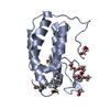 7rjlC  7rjmC  7rjoC  6qjuS S: Starting model for refinement C: citing same article ( |
|---|---|
| Similar structure data |
-Links
 PDBj
PDBj- Assembly
| Deposited unit | 
| ||||||||
|---|---|---|---|---|---|---|---|---|---|
| 1 | 
| ||||||||
| 2 | 
| ||||||||
| Unit cell |
|
-Components
| #1: Protein | Mass: 14586.843 Da / Num. of mol.: 2 Source method: isolated from a genetically manipulated source Source: (gene. exp.) Homo sapiens (human) / Gene: BRD3, KIAA0043, RING3L / Production host:  #2: Protein/peptide | Mass: 1235.475 Da / Num. of mol.: 2 / Fragment: UNP residues 330-339 / Source method: obtained synthetically / Source: (synth.) Homo sapiens (human) / References: UniProt: Q9NYF8#3: Chemical |   Mass: 62.068 Da / Num. of mol.: 3 / Source method: obtained synthetically / Formula: C2H6O2 Mass: 62.068 Da / Num. of mol.: 3 / Source method: obtained synthetically / Formula: C2H6O2#4: Water | ChemComp-HOH / |  Mass: 18.015 Da / Num. of mol.: 139 / Source method: isolated from a natural source / Formula: H2O Mass: 18.015 Da / Num. of mol.: 139 / Source method: isolated from a natural source / Formula: H2OHas ligand of interest | Y | Has protein modification | Y | |
|---|
-Experimental details
-Experiment
| Experiment | Method: X-RAY DIFFRACTION / Number of used crystals: 1 |
|---|
- Sample preparation
| Crystal | Density Matthews: 2.26 Å3/Da / Density % sol: 45.6 % |
|---|---|
| Crystal grow | Temperature: 292 K / Method: vapor diffusion, sitting drop / pH: 4.6 Details: 0.2 M ammonium acetate, 0.1 M sodium acetate trihydrate, 30% w/v PEG4000 |
-Data collection
| Diffraction | Mean temperature: 100 K / Serial crystal experiment: N | ||||||||||||||||||||||||||||||
|---|---|---|---|---|---|---|---|---|---|---|---|---|---|---|---|---|---|---|---|---|---|---|---|---|---|---|---|---|---|---|---|
| Diffraction source | Source: SYNCHROTRON / Site: NSLS-II / Beamline: 17-ID-1 / Wavelength: 0.92 Å | ||||||||||||||||||||||||||||||
| Detector | Type: DECTRIS EIGER X 16M / Detector: PIXEL / Date: Mar 31, 2020 | ||||||||||||||||||||||||||||||
| Radiation | Monochromator: double crystal Si(111) / Protocol: SINGLE WAVELENGTH / Monochromatic (M) / Laue (L): M / Scattering type: x-ray | ||||||||||||||||||||||||||||||
| Radiation wavelength | Wavelength: 0.92 Å / Relative weight: 1 | ||||||||||||||||||||||||||||||
| Reflection | Resolution: 1.95→29.49 Å / Num. obs: 21679 / % possible obs: 100 % / Redundancy: 13.2 % / CC1/2: 0.997 / Rmerge(I) obs: 0.133 / Rpim(I) all: 0.038 / Rrim(I) all: 0.139 / Net I/σ(I): 14.3 / Num. measured all: 285809 / Scaling rejects: 350 | ||||||||||||||||||||||||||||||
| Reflection shell | Diffraction-ID: 1
|
-Phasing
| Phasing | Method: molecular replacement |
|---|
- Processing
| Software |
| ||||||||||||||||||||||||
|---|---|---|---|---|---|---|---|---|---|---|---|---|---|---|---|---|---|---|---|---|---|---|---|---|---|
| Refinement | Method to determine structure: MOLECULAR REPLACEMENT Starting model: PDB entry 6QJU Resolution: 1.95→20 Å / Cross valid method: THROUGHOUT
| ||||||||||||||||||||||||
| Displacement parameters | Biso mean: 25.4 Å2 | ||||||||||||||||||||||||
| Refinement step | Cycle: LAST / Resolution: 1.95→20 Å
| ||||||||||||||||||||||||
| LS refinement shell | Resolution: 1.95→2.02 Å / Rfactor Rfree error: 0
|
