 Movie
Movie Controller

[English] 日本語
 Yorodumi
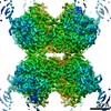
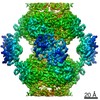
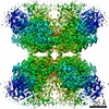
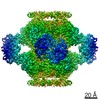
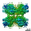
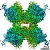
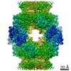
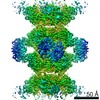
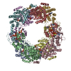
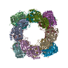
- PDB-7rfh: HUMAN RETINAL VARIANT IMPDH1(595) TREATED WITH ATP, OCTAMER-CENTERED -
+ Open data

- Basic information
| Entry | Database: PDB / ID: 7rfh | ||||||||||||
|---|---|---|---|---|---|---|---|---|---|---|---|---|---|
| Title | HUMAN RETINAL VARIANT IMPDH1(595) TREATED WITH ATP, OCTAMER-CENTERED | ||||||||||||
 Components Components | Isoform 5 of Inosine-5'-monophosphate dehydrogenase 1 | ||||||||||||
 Keywords Keywords | OXIDOREDUCTASE / METABOLISM / FILAMENT / ALLOSTERY / ADENINE | ||||||||||||
| Function / homology | IMP dehydrogenase / ADENOSINE-5'-TRIPHOSPHATE / Isoform 5 of Inosine-5'-monophosphate dehydrogenase 1 Function and homology information Function and homology information | ||||||||||||
| Biological species |  Homo sapiens (human) Homo sapiens (human) | ||||||||||||
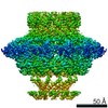
| Method | ELECTRON MICROSCOPY / single particle reconstruction / cryo EM / Resolution: 3.7 Å | ||||||||||||
| Model details | FILAMENT ASSEMBLY INTERFACE RECONSTRUCTION | ||||||||||||
 Authors Authors | Burrell, A.L. / Kollman, J.M. | ||||||||||||
| Funding support |  United States, 3items United States, 3items
| ||||||||||||
 Citation Citation | Journal: Nat Struct Mol Biol / Year: 2022 Title: IMPDH1 retinal variants control filament architecture to tune allosteric regulation. Authors: Anika L Burrell / Chuankai Nie / Meerit Said / Jacqueline C Simonet / David Fernández-Justel / Matthew C Johnson / Joel Quispe / Rubén M Buey / Jeffrey R Peterson / Justin M Kollman /  Abstract: Inosine-5'-monophosphate dehydrogenase (IMPDH), a key regulatory enzyme in purine nucleotide biosynthesis, dynamically assembles filaments in response to changes in metabolic demand. Humans have two ...Inosine-5'-monophosphate dehydrogenase (IMPDH), a key regulatory enzyme in purine nucleotide biosynthesis, dynamically assembles filaments in response to changes in metabolic demand. Humans have two isoforms: IMPDH2 filaments reduce sensitivity to feedback inhibition, while IMPDH1 assembly remains uncharacterized. IMPDH1 plays a unique role in retinal metabolism, and point mutants cause blindness. Here, in a series of cryogenic-electron microscopy structures we show that human IMPDH1 assembles polymorphic filaments with different assembly interfaces in extended and compressed states. Retina-specific splice variants introduce structural elements that reduce sensitivity to GTP inhibition, including stabilization of the extended filament form. Finally, we show that IMPDH1 disease mutations fall into two classes: one disrupts GTP regulation and the other has no effect on GTP regulation or filament assembly. These findings provide a foundation for understanding the role of IMPDH1 in retinal function and disease and demonstrate the diverse mechanisms by which metabolic enzyme filaments are allosterically regulated. | ||||||||||||
| History |
|
- Structure visualization
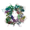
| Movie |
Movie viewer |
|---|---|
| Structure viewer | Molecule: MolmilJmol/JSmol |
- Downloads & links
-Download
| PDBx/mmCIF format | 7rfh.cif.gz | 1.1 MB | Display | PDBx/mmCIF format |
|---|---|---|---|---|
| PDB format | pdb7rfh.ent.gz | 960 KB | Display | PDB format |
| PDBx/mmJSON format | 7rfh.json.gz | Tree view | PDBx/mmJSON format | |
| Others |  Other downloads Other downloads |
-Validation report
| Arichive directory | https://data.pdbj.org/pub/pdb/validation_reports/rf/7rfhftp://data.pdbj.org/pub/pdb/validation_reports/rf/7rfh | HTTPS FTP |
|---|
-Related structure data
| Related structure data |  24442MC 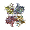 7rerC 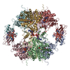 7resC  7rfeC  7rffC 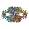 7rfgC 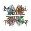 7rfiC 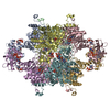 7rgdC 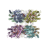 7rgiC 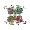 7rglC 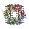 7rgmC  7rgqC C: citing same article ( M: map data used to model this data |
|---|---|
| Similar structure data |


-Links
 PDBj
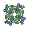
PDBj- Assembly
| Deposited unit | 
|
|---|---|
| 1 |
|
-Components
| #1: Protein | Mass: 63407.324 Da / Num. of mol.: 8 Source method: isolated from a genetically manipulated source Source: (gene. exp.) Homo sapiens (human) / Gene: IMPDH1, IMPD1 / Plasmid: PSMT3 / Production host:  #2: Chemical | ChemComp-ATP /   Mass: 507.181 Da / Num. of mol.: 16 / Source method: obtained synthetically / Formula: C10H16N5O13P3 / Feature type: SUBJECT OF INVESTIGATION / Comment: ATP, energy-carrying molecule*YM Mass: 507.181 Da / Num. of mol.: 16 / Source method: obtained synthetically / Formula: C10H16N5O13P3 / Feature type: SUBJECT OF INVESTIGATION / Comment: ATP, energy-carrying molecule*YMHas ligand of interest | Y | |
|---|
-Experimental details
-Experiment
| Experiment | Method: ELECTRON MICROSCOPY |
|---|---|
| EM experiment | Aggregation state: FILAMENT / 3D reconstruction method: single particle reconstruction |
- Sample preparation
| Component | Name: Assembly interface of IMPDH1 filament bound to ATP, IMP, NAD+ Type: COMPLEX / Entity ID: #1 / Source: RECOMBINANT |
|---|---|
| Molecular weight | Value: 55405 MDa / Experimental value: NO |
| Source (natural) | Organism: Homo sapiens (human) |
| Source (recombinant) | Organism: |
| Buffer solution | pH: 7 |
| Specimen | Embedding applied: NO / Shadowing applied: NO / Staining applied: NO / Vitrification applied: YES |
| Vitrification | Cryogen name: ETHANE |
- Electron microscopy imaging
| Experimental equipment |  Model: Titan Krios / Image courtesy: FEI Company |
|---|---|
| Microscopy | Model: FEI TITAN KRIOS |
| Electron gun | Electron source:  FIELD EMISSION GUN / Accelerating voltage: 300 kV / Illumination mode: FLOOD BEAM FIELD EMISSION GUN / Accelerating voltage: 300 kV / Illumination mode: FLOOD BEAM |
| Electron lens | Mode: BRIGHT FIELD |
| Image recording | Electron dose: 50 e/Å2 / Film or detector model: GATAN K2 SUMMIT (4k x 4k) |
- Processing
| CTF correction | Type: PHASE FLIPPING AND AMPLITUDE CORRECTION |
|---|---|
| 3D reconstruction | Resolution: 3.7 Å / Resolution method: FSC 0.143 CUT-OFF / Num. of particles: 11476 / Symmetry type: POINT |
