- PDB-4dqw: Crystal Structure Analysis of PA3770 -
+
Open data
ID or keywords:
Loading...
-
Basic information
Entry
Database: PDB / ID: 4dqw
Title
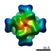
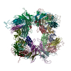
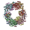
Crystal Structure Analysis of PA3770
Components
Inosine-5'-monophosphate dehydrogenase
Keywords
OXIDOREDUCTASE / IMPDH enzyme
Function / homology
Function and homology information
IMP dehydrogenase / IMP dehydrogenase activity / GMP biosynthetic process / GTP biosynthetic process / ATP binding / metal ion binding / identical protein binding Similarity search - Function
CBS-domain / CBS-domain / IMP dehydrogenase / GMP reductase, conserved site / IMP dehydrogenase / GMP reductase signature. / Inosine-5'-monophosphate dehydrogenase / IMP dehydrogenase/GMP reductase / IMP dehydrogenase / GMP reductase domain / IMP dehydrogenase / GMP reductase domain / Domain in cystathionine beta-synthase and other proteins. / CBS domain superfamily ...CBS-domain / CBS-domain / IMP dehydrogenase / GMP reductase, conserved site / IMP dehydrogenase / GMP reductase signature. / Inosine-5'-monophosphate dehydrogenase / IMP dehydrogenase/GMP reductase / IMP dehydrogenase / GMP reductase domain / IMP dehydrogenase / GMP reductase domain / Domain in cystathionine beta-synthase and other proteins. / CBS domain superfamily / CBS domain / CBS domain / CBS domain profile. / Aldolase class I / Aldolase-type TIM barrel / TIM Barrel / Alpha-Beta Barrel / Roll / Alpha Beta Similarity search - Domain/homology
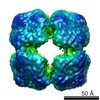
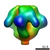
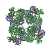
Journal: Structure / Year: 2013 Title: MgATP regulates allostery and fiber formation in IMPDHs. Authors: Gilles Labesse / Thomas Alexandre / Laurène Vaupré / Isabelle Salard-Arnaud / Joséphine Lai Kee Him / Bertrand Raynal / Patrick Bron / Hélène Munier-Lehmann / Abstract: Inosine-5'-monophosphate dehydrogenase (IMPDH) is a rate-limiting enzyme in nucleotide biosynthesis studied as an important therapeutic target and its complex functioning in vivo is still puzzling ...Inosine-5'-monophosphate dehydrogenase (IMPDH) is a rate-limiting enzyme in nucleotide biosynthesis studied as an important therapeutic target and its complex functioning in vivo is still puzzling and debated. Here, we highlight the structural basis for the regulation of IMPDHs by MgATP. Our results demonstrate the essential role of the CBS tandem, conserved among almost all IMPDHs. We found that Pseudomonas aeruginosa IMPDH is an octameric enzyme allosterically regulated by MgATP and showed that this octameric organization is widely conserved in the crystal structures of other IMPDHs. We also demonstrated that human IMPDH1 adopts two types of complementary octamers that can pile up into isolated fibers in the presence of MgATP. The aggregation of such fibers in the autosomal dominant mutant, D226N, could explain the onset of the retinopathy adRP10. Thus, the regulatory CBS modules in IMPDHs are functional and they can either modulate catalysis or macromolecular assembly.
In the structure databanks used in Yorodumi, some data are registered as the other names, "COVID-19 virus" and "2019-nCoV". Here are the details of the virus and the list of structure data.
Jan 31, 2019. EMDB accession codes are about to change! (news from PDBe EMDB page)
EMDB accession codes are about to change! (news from PDBe EMDB page)
The allocation of 4 digits for EMDB accession codes will soon come to an end. Whilst these codes will remain in use, new EMDB accession codes will include an additional digit and will expand incrementally as the available range of codes is exhausted. The current 4-digit format prefixed with “EMD-” (i.e. EMD-XXXX) will advance to a 5-digit format (i.e. EMD-XXXXX), and so on. It is currently estimated that the 4-digit codes will be depleted around Spring 2019, at which point the 5-digit format will come into force.
The EM Navigator/Yorodumi systems omit the EMD- prefix.
Related info.:Q: What is EMD? / ID/Accession-code notation in Yorodumi/EM Navigator
Yorodumi is a browser for structure data from EMDB, PDB, SASBDB, etc.
This page is also the successor to EM Navigator detail page, and also detail information page/front-end page for Omokage search.
The word "yorodu" (or yorozu) is an old Japanese word meaning "ten thousand". "mi" (miru) is to see.
Related info.:EMDB / PDB / SASBDB / Comparison of 3 databanks / Yorodumi Search / Aug 31, 2016. New EM Navigator & Yorodumi / Yorodumi Papers / Jmol/JSmol / Function and homology information / Changes in new EM Navigator and Yorodumi
 Movie
Movie Controller


 Open data
 Basic information
 Components
 Keywords
 Function and homology information
 Pseudomonas aeruginosa (bacteria)
 X-RAY DIFFRACTION /
  Authors
 Citation
 Structure visualization
 Downloads & links
 Other downloads
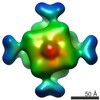

 PDBj
PDBj
 Assembly


 Mass: 507.181 Da / Num. of mol.: 4 / Source method: obtained synthetically / Formula: C10H16N5O13P3 / Comment: ATP, energy-carrying molecule*YM
 Mass: 54.938 Da / Num. of mol.: 4 / Source method: obtained synthetically / Formula: Mn
 Mass: 94.971 Da / Num. of mol.: 1 / Source method: obtained synthetically / Formula: PO4
 Mass: 92.094 Da / Num. of mol.: 1 / Source method: obtained synthetically / Formula: C3H8O3
 Sample preparation
 Processing