 Movie
Movie Controller

[English] 日本語
 Yorodumi
- EMDB-5438: 3D reconstruction of a self-assembling designed oligomer with oct... -
+ Open data

- Basic information
| Entry | Database: EMDB / ID: EMD-5438 | |||||||||
|---|---|---|---|---|---|---|---|---|---|---|
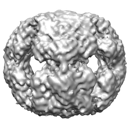
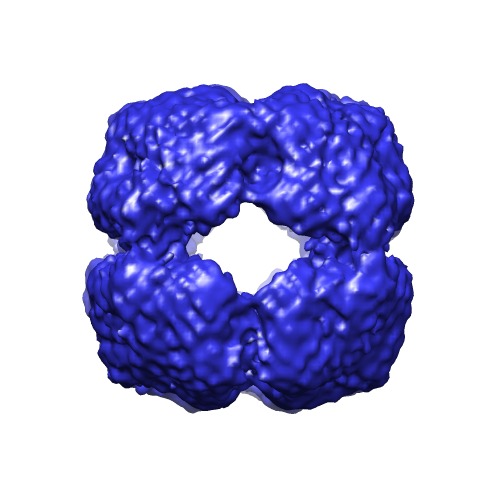
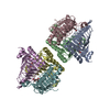
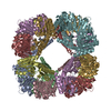
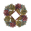
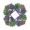
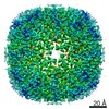
| Title | 3D reconstruction of a self-assembling designed oligomer with octahedral symmetry | |||||||||
 Map data Map data | Reconstruction of designed self-assembling protein oligomer with octahedral symmetry | |||||||||
 Sample Sample |
| |||||||||
 Keywords Keywords | octahedral symmetry / designed | |||||||||
| Function / homology |  Function and homology information Function and homology informationpropanediol degradation polyhedral organelle / 1,2-propanediol catabolic process / 4 iron, 4 sulfur cluster binding / metal ion binding Similarity search - Function | |||||||||
| Biological species |  Salmonella enterica (bacteria) Salmonella enterica (bacteria) | |||||||||
| Method | single particle reconstruction / cryo EM / Resolution: 20.0 Å | |||||||||
 Authors Authors | Vollmar BS / King NP / Baker D / Gonen T | |||||||||
 Citation Citation | Journal: Science / Year: 2012 Title: Computational design of self-assembling protein nanomaterials with atomic level accuracy. Authors: Neil P King / William Sheffler / Michael R Sawaya / Breanna S Vollmar / John P Sumida / Ingemar André / Tamir Gonen / Todd O Yeates / David Baker /  Abstract: We describe a general computational method for designing proteins that self-assemble to a desired symmetric architecture. Protein building blocks are docked together symmetrically to identify ...We describe a general computational method for designing proteins that self-assemble to a desired symmetric architecture. Protein building blocks are docked together symmetrically to identify complementary packing arrangements, and low-energy protein-protein interfaces are then designed between the building blocks in order to drive self-assembly. We used trimeric protein building blocks to design a 24-subunit, 13-nm diameter complex with octahedral symmetry and a 12-subunit, 11-nm diameter complex with tetrahedral symmetry. The designed proteins assembled to the desired oligomeric states in solution, and the crystal structures of the complexes revealed that the resulting materials closely match the design models. The method can be used to design a wide variety of self-assembling protein nanomaterials. | |||||||||
| History |
|
- Structure visualization
| Movie |
Movie viewer |
|---|---|
| Structure viewer | EM map: SurfViewMolmilJmol/JSmol |
| Supplemental images |
- Downloads & links
-EMDB archive
| Map data | emd_5438.map.gz | 2.2 MB | EMDB map data format | |
|---|---|---|---|---|
| Header (meta data) | emd-5438-v30.xmlemd-5438.xml | 9.5 KB 9.5 KB | Display Display | EMDB header |
| Images |  emd_5438_1.jpgemd_5438_2.tif emd_5438_1.jpgemd_5438_2.tif | 49.5 KB 178.5 KB | ||
| Archive directory |  http://ftp.pdbj.org/pub/emdb/structures/EMD-5438ftp://ftp.pdbj.org/pub/emdb/structures/EMD-5438 http://ftp.pdbj.org/pub/emdb/structures/EMD-5438ftp://ftp.pdbj.org/pub/emdb/structures/EMD-5438 | HTTPS FTP |
-Related structure data


-Links
| EMDB pages | EMDB (EBI/PDBe) / EMDataResource |
|---|---|
| Related items in Molecule of the Month |


-Map
| File | Download / File: emd_5438.map.gz / Format: CCP4 / Size: 6.4 MB / Type: IMAGE STORED AS FLOATING POINT NUMBER (4 BYTES) | ||||||||||||||||||||||||||||||||||||||||||||||||||||||||||||||||||||
|---|---|---|---|---|---|---|---|---|---|---|---|---|---|---|---|---|---|---|---|---|---|---|---|---|---|---|---|---|---|---|---|---|---|---|---|---|---|---|---|---|---|---|---|---|---|---|---|---|---|---|---|---|---|---|---|---|---|---|---|---|---|---|---|---|---|---|---|---|---|
| Annotation | Reconstruction of designed self-assembling protein oligomer with octahedral symmetry | ||||||||||||||||||||||||||||||||||||||||||||||||||||||||||||||||||||
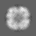
| Projections & slices | Image control
Images are generated by Spider. | ||||||||||||||||||||||||||||||||||||||||||||||||||||||||||||||||||||
| Voxel size | X=Y=Z: 1.474 Å | ||||||||||||||||||||||||||||||||||||||||||||||||||||||||||||||||||||
| Density |
| ||||||||||||||||||||||||||||||||||||||||||||||||||||||||||||||||||||
| Symmetry | Space group: 1 | ||||||||||||||||||||||||||||||||||||||||||||||||||||||||||||||||||||
| Details | EMDB XML:
CCP4 map header:
| ||||||||||||||||||||||||||||||||||||||||||||||||||||||||||||||||||||
 Z (Sec.)
 Y (Row.)
 X (Col.)


-Supplemental data
- Sample components
-Entire : Designed protein oligomer with octahedral symmetry. The model for...
| Entire | Name: Designed protein oligomer with octahedral symmetry. The model for the design is PduT from Salmonella enterica with the following mutations: K15A, C38S, M67L, N148A, N149L, E156S, E160A, K161Y, R167A, V169L |
|---|---|
| Components |
|
-Supramolecule #1000: Designed protein oligomer with octahedral symmetry. The model for...
| Supramolecule | Name: Designed protein oligomer with octahedral symmetry. The model for the design is PduT from Salmonella enterica with the following mutations: K15A, C38S, M67L, N148A, N149L, E156S, E160A, K161Y, R167A, V169L type: sample / ID: 1000 / Details: Monodisperse sample / Oligomeric state: 24 / Number unique components: 1 |
|---|---|
| Molecular weight | Theoretical: 480 KDa |
-Macromolecule #1: Propanediol utilization polyhedral body protein PduT
| Macromolecule | Name: Propanediol utilization polyhedral body protein PduT / type: protein_or_peptide / ID: 1 Details: Mutations: K15A, C38S, M67L, N148A, N149L, E156S, E160A, K161Y, R167A, V169L Number of copies: 24 / Recombinant expression: Yes |
|---|---|
| Source (natural) | Organism: Salmonella enterica (bacteria) |
| Recombinant expression | Organism: |
-Experimental details
-Structure determination
| Method | cryo EM |
|---|---|
 Processing Processing | single particle reconstruction |
| Aggregation state | particle |
-Sample preparation
| Concentration | 0.3 mg/mL |
|---|---|
| Buffer | pH: 8 / Details: 25 mM Tris, pH 8.0, 150 mM NaCl, 1 mM DTT |
| Grid | Details: Quantifoil R1.2/1.3 holey carbon 400 mesh copper grids |
| Vitrification | Cryogen name: ETHANE / Chamber humidity: 100 % / Instrument: FEI VITROBOT MARK III Method: Blotted with filter paper and plunged into liquid ethane. |
- Electron microscopy
| Microscope | FEI TECNAI F20 |
|---|---|
| Date | Jul 12, 2011 |
| Image recording | Category: CCD / Film or detector model: TVIPS TEMCAM-F816 (8k x 8k) / Number real images: 565 |
| Tilt angle min | 0 |
| Tilt angle max | 0 |
| Electron beam | Acceleration voltage: 200 kV / Electron source:  FIELD EMISSION GUN FIELD EMISSION GUN |
| Electron optics | Calibrated magnification: 217096 / Illumination mode: SPOT SCAN / Imaging mode: BRIGHT FIELD / Cs: 2.12 mm / Nominal defocus max: 2.0 µm / Nominal defocus min: 1.0 µm / Nominal magnification: 100000 |
| Sample stage | Specimen holder model: GATAN LIQUID NITROGEN |
| Experimental equipment |  Model: Tecnai F20 / Image courtesy: FEI Company |
-Image processing
| Details | Particles were selected using the automatic selection program Electron Micrograph Utility (cryoem.ucsf.edu). |
|---|---|
| CTF correction | Details: CTFFIND3 |
| Final reconstruction | Resolution.type: BY AUTHOR / Resolution: 20.0 Å / Resolution method: FSC 0.5 CUT-OFF / Software - Name: FREALIGN Details: Reconstruction was calculated based on 2x binned images yielding a pixel size of 1.474 A/pixel. Number images used: 42025 |
