 Movie
Movie Controller


+ Open data

- Basic information
| Entry | Database: PDB / ID: 7rew | ||||||
|---|---|---|---|---|---|---|---|
| Title | Crystal Structure of IL-13 in complex with MMAb3 Fab | ||||||
 Components Components |
| ||||||
 Keywords Keywords | CYTOKINE / alpha-helical bundle / protein-Fab complex | ||||||
| Function / homology |  Function and homology information Function and homology informationcytokine receptor binding / macrophage activation / cytokine activity / immune response / extracellular space Similarity search - Function | ||||||
| Biological species |  Homo sapiens (human) Homo sapiens (human) | ||||||
| Method |  X-RAY DIFFRACTION / SYNCHROTRON / MOLECULAR REPLACEMENT / Resolution: 2.1 Å X-RAY DIFFRACTION / SYNCHROTRON / MOLECULAR REPLACEMENT / Resolution: 2.1 Å | ||||||
| Model details | Fc | ||||||
 Authors Authors | Sudom, A. / Min, X. | ||||||
 Citation Citation | Journal: J.Biol.Chem. / Year: 2022 Title: Development of a potent high-affinity human therapeutic antibody via novel application of recombination signal sequence-based affinity maturation. Authors: Kielczewska, A. / D'Angelo, I. / Amador, M.S. / Wang, T. / Sudom, A. / Min, X. / Rathanaswami, P. / Pigott, C. / Foltz, I.N. | ||||||
| History |
|
- Structure visualization
| Structure viewer | Molecule: MolmilJmol/JSmol |
|---|
- Downloads & links
-Download
| PDBx/mmCIF format | 7rew.cif.gz | 248.8 KB | Display | PDBx/mmCIF format |
|---|---|---|---|---|
| PDB format | pdb7rew.ent.gz | 160.4 KB | Display | PDB format |
| PDBx/mmJSON format | 7rew.json.gz | Tree view | PDBx/mmJSON format | |
| Others |  Other downloads Other downloads |
-Validation report
| Arichive directory | https://data.pdbj.org/pub/pdb/validation_reports/re/7rewftp://data.pdbj.org/pub/pdb/validation_reports/re/7rew | HTTPS FTP |
|---|
-Related structure data
| Related structure data | 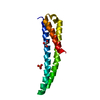 4cqiS S: Starting model for refinement |
|---|---|
| Similar structure data |
-Links
 PDBj
PDBj


- Assembly
| Deposited unit | 
| ||||||||||
|---|---|---|---|---|---|---|---|---|---|---|---|
| 1 | 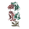
| ||||||||||
| 2 | 
| ||||||||||
| Unit cell |
|
-Components
| #1: Antibody | Mass: 24108.861 Da / Num. of mol.: 2 Source method: isolated from a genetically manipulated source Source: (gene. exp.) Homo sapiens (human) / Cell line (production host): 293T / Production host: Homo sapiens (human) / Tissue (production host): kidney#2: Antibody | Mass: 22737.080 Da / Num. of mol.: 2 Source method: isolated from a genetically manipulated source Source: (gene. exp.) Homo sapiens (human) / Cell line (production host): 293T / Production host: Homo sapiens (human) / Tissue (production host): kidney#3: Protein | Mass: 12302.288 Da / Num. of mol.: 2 / Fragment: UNP residues 22-132 Source method: isolated from a genetically manipulated source Source: (gene. exp.) Gene: IL13 / Cell line (production host): 293T / Production host: Homo sapiens (human) / Tissue (production host): kidney / References: UniProt: Q0PW92#4: Water | ChemComp-HOH / |  Mass: 18.015 Da / Num. of mol.: 131 / Source method: isolated from a natural source / Formula: H2O Mass: 18.015 Da / Num. of mol.: 131 / Source method: isolated from a natural source / Formula: H2OHas protein modification | Y | |
|---|
-Experimental details
-Experiment
| Experiment | Method: X-RAY DIFFRACTION / Number of used crystals: 1 |
|---|
- Sample preparation
| Crystal | Density Matthews: 2.31 Å3/Da / Density % sol: 46.83 % / Mosaicity: 0.3 ° |
|---|---|
| Crystal grow | Temperature: 293 K / Method: evaporation / pH: 8.5 Details: 0.2 M LiSO4, 0.1 M Tris HCl pH 8.5, 25% PEG 5,000 MME |
-Data collection
| Diffraction | Mean temperature: 100 K / Serial crystal experiment: N | |||||||||||||||||||||||||||
|---|---|---|---|---|---|---|---|---|---|---|---|---|---|---|---|---|---|---|---|---|---|---|---|---|---|---|---|---|
| Diffraction source | Source: SYNCHROTRON / Site: APS  / Beamline: 22-ID / Wavelength: 1 Å / Beamline: 22-ID / Wavelength: 1 Å | |||||||||||||||||||||||||||
| Detector | Type: RAYONIX MX300-HS / Detector: CCD / Date: Apr 4, 2018 | |||||||||||||||||||||||||||
| Radiation | Monochromator: Si / Protocol: SINGLE WAVELENGTH / Monochromatic (M) / Laue (L): M / Scattering type: x-ray | |||||||||||||||||||||||||||
| Radiation wavelength | Wavelength: 1 Å / Relative weight: 1 | |||||||||||||||||||||||||||
| Reflection | Resolution: 2.1→68.94 Å / Num. obs: 54189 / % possible obs: 88 % / Redundancy: 2.1 % / Biso Wilson estimate: 39.04 Å2 / CC1/2: 0.993 / Rmerge(I) obs: 0.051 / Rpim(I) all: 0.044 / Rrim(I) all: 0.068 / Net I/σ(I): 7.2 / Num. measured all: 115864 / Scaling rejects: 418 | |||||||||||||||||||||||||||
| Reflection shell | Diffraction-ID: 1 / Redundancy: 2.1 %
|
- Processing
| Software |
| ||||||||||||||||||||||||||||||||||||||||||||||||||||||||||||||||||||||||||||||||||||||||||||||||||||||||||||||||||||||||||||||||||||||||||||
|---|---|---|---|---|---|---|---|---|---|---|---|---|---|---|---|---|---|---|---|---|---|---|---|---|---|---|---|---|---|---|---|---|---|---|---|---|---|---|---|---|---|---|---|---|---|---|---|---|---|---|---|---|---|---|---|---|---|---|---|---|---|---|---|---|---|---|---|---|---|---|---|---|---|---|---|---|---|---|---|---|---|---|---|---|---|---|---|---|---|---|---|---|---|---|---|---|---|---|---|---|---|---|---|---|---|---|---|---|---|---|---|---|---|---|---|---|---|---|---|---|---|---|---|---|---|---|---|---|---|---|---|---|---|---|---|---|---|---|---|---|---|
| Refinement | Method to determine structure: MOLECULAR REPLACEMENT Starting model: 4CQI Resolution: 2.1→35.96 Å / SU ML: 0.3344 / Cross valid method: THROUGHOUT / σ(F): 1.97 / Phase error: 36.4314 / Stereochemistry target values: CDL v1.2
| ||||||||||||||||||||||||||||||||||||||||||||||||||||||||||||||||||||||||||||||||||||||||||||||||||||||||||||||||||||||||||||||||||||||||||||
| Solvent computation | Shrinkage radii: 0.9 Å / VDW probe radii: 1.11 Å / Solvent model: FLAT BULK SOLVENT MODEL | ||||||||||||||||||||||||||||||||||||||||||||||||||||||||||||||||||||||||||||||||||||||||||||||||||||||||||||||||||||||||||||||||||||||||||||
| Displacement parameters | Biso mean: 52.78 Å2 | ||||||||||||||||||||||||||||||||||||||||||||||||||||||||||||||||||||||||||||||||||||||||||||||||||||||||||||||||||||||||||||||||||||||||||||
| Refinement step | Cycle: LAST / Resolution: 2.1→35.96 Å
| ||||||||||||||||||||||||||||||||||||||||||||||||||||||||||||||||||||||||||||||||||||||||||||||||||||||||||||||||||||||||||||||||||||||||||||
| Refine LS restraints |
| ||||||||||||||||||||||||||||||||||||||||||||||||||||||||||||||||||||||||||||||||||||||||||||||||||||||||||||||||||||||||||||||||||||||||||||
| LS refinement shell |
|
