 Movie
Movie Controller

[English] 日本語
 Yorodumi
- PDB-7rcz: Crystal structure of C. difficile SpoVD in complex with ampicillin -
+ Open data

- Basic information
| Entry | Database: PDB / ID: 7rcz | |||||||||||||||
|---|---|---|---|---|---|---|---|---|---|---|---|---|---|---|---|---|
| Title | Crystal structure of C. difficile SpoVD in complex with ampicillin | |||||||||||||||
 Components Components | Stage V sporulation protein D (Sporulation specific penicillin-binding protein) | |||||||||||||||
 Keywords Keywords | PEPTIDE BINDING PROTEIN / Peptidoglycan / PBP / PBP2 / cell wall / transpeptidase / b-lactam / gram-positive / spore / sporulation | |||||||||||||||
| Function / homology | DI(HYDROXYETHYL)ETHER / N-PROPANOL / Chem-ZZ7 / :  Function and homology information Function and homology information | |||||||||||||||
| Biological species |  Clostridioides difficile (bacteria) Clostridioides difficile (bacteria) | |||||||||||||||
| Method |  X-RAY DIFFRACTION / SYNCHROTRON / MOLECULAR REPLACEMENT / Resolution: 2.2 Å X-RAY DIFFRACTION / SYNCHROTRON / MOLECULAR REPLACEMENT / Resolution: 2.2 Å | |||||||||||||||
 Authors Authors | Sacco, M. / Chen, Y. | |||||||||||||||
| Funding support |  United States, 4items United States, 4items
| |||||||||||||||
 Citation Citation | Journal: Nat Commun / Year: 2022 Title: A unique class of Zn 2+ -binding serine-based PBPs underlies cephalosporin resistance and sporogenesis in Clostridioides difficile. Authors: Sacco, M.D. / Wang, S. / Adapa, S.R. / Zhang, X. / Lewandowski, E.M. / Gongora, M.V. / Keramisanou, D. / Atlas, Z.D. / Townsend, J.A. / Gatdula, J.R. / Morgan, R.T. / Hammond, L.R. / Marty, ...Authors: Sacco, M.D. / Wang, S. / Adapa, S.R. / Zhang, X. / Lewandowski, E.M. / Gongora, M.V. / Keramisanou, D. / Atlas, Z.D. / Townsend, J.A. / Gatdula, J.R. / Morgan, R.T. / Hammond, L.R. / Marty, M.T. / Wang, J. / Eswara, P.J. / Gelis, I. / Jiang, R.H.Y. / Sun, X. / Chen, Y. | |||||||||||||||
| History |
|
- Structure visualization
| Structure viewer | Molecule: MolmilJmol/JSmol |
|---|
- Downloads & links
-Download
| PDBx/mmCIF format | 7rcz.cif.gz | 223.3 KB | Display | PDBx/mmCIF format |
|---|---|---|---|---|
| PDB format | pdb7rcz.ent.gz | 174.4 KB | Display | PDB format |
| PDBx/mmJSON format | 7rcz.json.gz | Tree view | PDBx/mmJSON format | |
| Others |  Other downloads Other downloads |
-Validation report
| Arichive directory | https://data.pdbj.org/pub/pdb/validation_reports/rc/7rczftp://data.pdbj.org/pub/pdb/validation_reports/rc/7rcz | HTTPS FTP |
|---|
-Related structure data
| Related structure data |  7rcwC 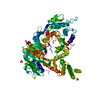 7rcxC 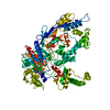 7rcyC 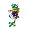 7rd0C 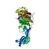 6un1S S: Starting model for refinement C: citing same article ( |
|---|---|
| Similar structure data |
-Links
 PDBj
PDBj
- Assembly
| Deposited unit | 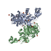
| ||||||||
|---|---|---|---|---|---|---|---|---|---|
| 1 |
| ||||||||
| Unit cell |
|
-Components
-Protein , 1 types, 2 molecules AB
| #1: Protein | Mass: 60601.922 Da / Num. of mol.: 2 Source method: isolated from a genetically manipulated source Source: (gene. exp.) Clostridioides difficile (strain R20291) (bacteria)Strain: R20291 / Gene: spoVD, CDR20291_2544 Production host: References: UniProt: C9YPN0 |
|---|
-Non-polymers , 7 types, 336 molecules 


| #2: Chemical | ChemComp-SO4 /  Mass: 96.063 Da / Num. of mol.: 10 / Source method: obtained synthetically / Formula: SO4 Mass: 96.063 Da / Num. of mol.: 10 / Source method: obtained synthetically / Formula: SO4#3: Chemical |  Mass: 238.305 Da / Num. of mol.: 2 / Source method: obtained synthetically / Formula: C8H18N2O4S / Comment: pH buffer*YM Mass: 238.305 Da / Num. of mol.: 2 / Source method: obtained synthetically / Formula: C8H18N2O4S / Comment: pH buffer*YM#4: Chemical | ChemComp-POL / |  Mass: 60.095 Da / Num. of mol.: 1 / Source method: obtained synthetically / Formula: C3H8O Mass: 60.095 Da / Num. of mol.: 1 / Source method: obtained synthetically / Formula: C3H8O#5: Chemical | ChemComp-PEG / |  Mass: 106.120 Da / Num. of mol.: 1 / Source method: obtained synthetically / Formula: C4H10O3 Mass: 106.120 Da / Num. of mol.: 1 / Source method: obtained synthetically / Formula: C4H10O3#6: Chemical |  Mass: 65.409 Da / Num. of mol.: 2 / Source method: obtained synthetically / Formula: Zn Mass: 65.409 Da / Num. of mol.: 2 / Source method: obtained synthetically / Formula: Zn#7: Chemical |  Mass: 367.420 Da / Num. of mol.: 2 / Source method: obtained synthetically / Formula: C16H21N3O5S / Feature type: SUBJECT OF INVESTIGATION Mass: 367.420 Da / Num. of mol.: 2 / Source method: obtained synthetically / Formula: C16H21N3O5S / Feature type: SUBJECT OF INVESTIGATION#8: Water | ChemComp-HOH / | Mass: 18.015 Da / Num. of mol.: 318 / Source method: isolated from a natural source / Formula: H2O |
|---|
-Details
| Has ligand of interest | Y |
|---|---|
| Has protein modification | Y |
-Experimental details
-Experiment
| Experiment | Method: X-RAY DIFFRACTION / Number of used crystals: 1 |
|---|
- Sample preparation
| Crystal | Density Matthews: 2.69 Å3/Da / Density % sol: 54.19 % |
|---|---|
| Crystal grow | Temperature: 293 K / Method: vapor diffusion, hanging drop Details: 15 % PEG 3350, 0.2 M AmSO4, 10 % PropOH, 0.1 M Na Citrate pH 5.6 |
-Data collection
| Diffraction | Mean temperature: 100 K / Serial crystal experiment: N | ||||||||||||||||||||||||||||||
|---|---|---|---|---|---|---|---|---|---|---|---|---|---|---|---|---|---|---|---|---|---|---|---|---|---|---|---|---|---|---|---|
| Diffraction source | Source: SYNCHROTRON / Site: APS / Beamline: 19-ID / Wavelength: 0.97911 Å | ||||||||||||||||||||||||||||||
| Detector | Type: DECTRIS PILATUS3 6M / Detector: PIXEL / Date: Nov 21, 2020 | ||||||||||||||||||||||||||||||
| Radiation | Protocol: SINGLE WAVELENGTH / Monochromatic (M) / Laue (L): M / Scattering type: x-ray | ||||||||||||||||||||||||||||||
| Radiation wavelength | Wavelength: 0.97911 Å / Relative weight: 1 | ||||||||||||||||||||||||||||||
| Reflection | Resolution: 2.2→49.58 Å / Num. obs: 62415 / % possible obs: 94.6 % / Redundancy: 3.5 % / CC1/2: 0.985 / Rmerge(I) obs: 0.105 / Rpim(I) all: 0.063 / Rrim(I) all: 0.123 / Net I/σ(I): 7.3 | ||||||||||||||||||||||||||||||
| Reflection shell | Diffraction-ID: 1
|
- Processing
| Software |
| ||||||||||||||||||||||||||||||||||||||||||||||||||||||||||||
|---|---|---|---|---|---|---|---|---|---|---|---|---|---|---|---|---|---|---|---|---|---|---|---|---|---|---|---|---|---|---|---|---|---|---|---|---|---|---|---|---|---|---|---|---|---|---|---|---|---|---|---|---|---|---|---|---|---|---|---|---|---|
| Refinement | Method to determine structure: MOLECULAR REPLACEMENT Starting model: 6UN1 Resolution: 2.2→49.58 Å / Cor.coef. Fo:Fc: 0.953 / Cor.coef. Fo:Fc free: 0.933 / SU B: 6.018 / SU ML: 0.148 / Cross valid method: THROUGHOUT / σ(F): 0 / ESU R: 0.247 / ESU R Free: 0.195 / Stereochemistry target values: MAXIMUM LIKELIHOOD Details: HYDROGENS HAVE BEEN ADDED IN THE RIDING POSITIONS U VALUES : REFINED INDIVIDUALLY
| ||||||||||||||||||||||||||||||||||||||||||||||||||||||||||||
| Solvent computation | Ion probe radii: 0.8 Å / Shrinkage radii: 0.8 Å / VDW probe radii: 1.2 Å / Solvent model: MASK | ||||||||||||||||||||||||||||||||||||||||||||||||||||||||||||
| Displacement parameters | Biso max: 142.75 Å2 / Biso mean: 41.966 Å2 / Biso min: 14.08 Å2
| ||||||||||||||||||||||||||||||||||||||||||||||||||||||||||||
| Refinement step | Cycle: final / Resolution: 2.2→49.58 Å
| ||||||||||||||||||||||||||||||||||||||||||||||||||||||||||||
| Refine LS restraints |
| ||||||||||||||||||||||||||||||||||||||||||||||||||||||||||||
| LS refinement shell | Resolution: 2.2→2.257 Å / Rfactor Rfree error: 0 / Total num. of bins used: 20
|
