 Movie
Movie Controller

[English] 日本語
 Yorodumi
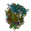
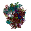
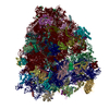
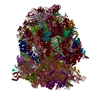
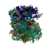
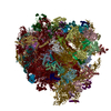
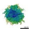
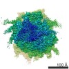
- PDB-7r81: Structure of the translating Neurospora crassa ribosome arrested ... -
+ Open data

- Basic information
| Entry | Database: PDB / ID: 7r81 | ||||||||||||
|---|---|---|---|---|---|---|---|---|---|---|---|---|---|
| Title | Structure of the translating Neurospora crassa ribosome arrested by cycloheximide | ||||||||||||
 Components Components |
| ||||||||||||
 Keywords Keywords | TRANSLATION / Protein Synthesis Inhibitor / Cytosolic Ribosome / Polysomal Ribosome / Peptidyl Transferase Center | ||||||||||||
| Function / homology |  Function and homology information Function and homology informationnegative regulation of cell integrity MAPK cascade / positive regulation of conjugation with cellular fusion / regulation of cytoplasmic translation / GCN2-mediated signaling / negative regulation of p38MAPK cascade / ribosome hibernation / preribosome / regulation of amino acid metabolic process / negative regulation of glucose mediated signaling pathway / positive regulation of translational fidelity ...negative regulation of cell integrity MAPK cascade / positive regulation of conjugation with cellular fusion / regulation of cytoplasmic translation / GCN2-mediated signaling / negative regulation of p38MAPK cascade / ribosome hibernation / preribosome / regulation of amino acid metabolic process / negative regulation of glucose mediated signaling pathway / positive regulation of translational fidelity / ribosome-associated ubiquitin-dependent protein catabolic process / pre-mRNA 5'-splice site binding / GDP-dissociation inhibitor activity / nonfunctional rRNA decay / preribosome, small subunit precursor / mRNA destabilization / negative regulation of mRNA splicing, via spliceosome / signaling receptor activator activity / negative regulation of translational frameshifting / translational elongation / G-protein alpha-subunit binding / endonucleolytic cleavage to generate mature 3'-end of SSU-rRNA from (SSU-rRNA, 5.8S rRNA, LSU-rRNA) / ribosomal subunit export from nucleus / protein-RNA complex assembly / endonucleolytic cleavage in ITS1 to separate SSU-rRNA from 5.8S rRNA and LSU-rRNA from tricistronic rRNA transcript (SSU-rRNA, 5.8S rRNA, LSU-rRNA) / protein-membrane adaptor activity / translation regulator activity / ribosomal small subunit export from nucleus / DNA-(apurinic or apyrimidinic site) endonuclease activity / positive regulation of autophagy / rescue of stalled cytosolic ribosome / protein kinase C binding / ribosomal large subunit biogenesis / maturation of LSU-rRNA from tricistronic rRNA transcript (SSU-rRNA, 5.8S rRNA, LSU-rRNA) / maturation of SSU-rRNA from tricistronic rRNA transcript (SSU-rRNA, 5.8S rRNA, LSU-rRNA) / maturation of SSU-rRNA / small-subunit processome / maintenance of translational fidelity / modification-dependent protein catabolic process / protein tag activity / rRNA processing / large ribosomal subunit / ribosomal small subunit assembly / ribosome biogenesis / ribosome binding / ribosomal small subunit biogenesis / 5S rRNA binding / ribosomal large subunit assembly / small ribosomal subunit / small ribosomal subunit rRNA binding / large ribosomal subunit rRNA binding / cytosolic small ribosomal subunit / cytosolic large ribosomal subunit / cytoplasmic translation / rRNA binding / structural constituent of ribosome / protein ubiquitination / ribosome / translation / G protein-coupled receptor signaling pathway / ribonucleoprotein complex / mRNA binding / ubiquitin protein ligase binding / nucleolus / RNA binding / zinc ion binding / nucleus / cytoplasm / cytosol Similarity search - Function | ||||||||||||
| Biological species |  Neurospora crassa (fungus) Neurospora crassa (fungus) | ||||||||||||
| Method | ELECTRON MICROSCOPY / single particle reconstruction / cryo EM / Resolution: 2.7 Å | ||||||||||||
 Authors Authors | Shen, L. / Su, Z. / Yang, K. / Wu, C. / Becker, T. / Bell-Pedersen, D. / Zhang, J. / Sachs, M.S. | ||||||||||||
| Funding support |  United States, 3items United States, 3items
| ||||||||||||
 Citation Citation | Journal: Proc Natl Acad Sci U S A / Year: 2021 Title: Structure of the translating ribosome arrested by cycloheximide. Authors: Lunda Shen / Zhaoming Su / Kailu Yang / Cheng Wu / Thomas Becker / Deborah Bell-Pedersen / Junjie Zhang / Matthew S Sachs /  Abstract: Ribosomes translate RNA into proteins. The protein synthesis inhibitor cycloheximide (CHX) is widely used to inhibit eukaryotic ribosomes engaged in translation elongation. However, the lack of ...Ribosomes translate RNA into proteins. The protein synthesis inhibitor cycloheximide (CHX) is widely used to inhibit eukaryotic ribosomes engaged in translation elongation. However, the lack of structural data for actively translating polyribosomes stalled by CHX leaves unanswered the question of which elongation step is inhibited. We elucidated CHX's mechanism of action based on the cryo-electron microscopy structure of actively translating ribosomes bound with CHX at 2.7-Å resolution. The ribosome structure from this filamentous fungus contains clearly resolved ribosomal protein eL28, like higher eukaryotes but unlike budding yeast, which lacks eL28. Despite some differences in overall structures, the ribosomes from , yeast, and humans all contain a highly conserved CHX binding site. We also sequenced classic CHX-resistant alleles. These mutations, including one at a residue not previously observed to affect CHX resistance in eukaryotes, were in the large subunit proteins uL15 and eL42 that are part of the CHX-binding pocket. In addition to A-site transfer RNA (tRNA), P-site tRNA, messenger RNA, and CHX that are associated with the translating ribosome, spermidine is present near the CHX binding site close to the E site on the large subunit. The tRNAs in the peptidyl transferase center are in the A/A site and the P/P site. The nascent peptide is attached to the A-site tRNA and not to the P-site tRNA. The structural and functional data obtained show that CHX arrests the ribosome in the classical PRE translocation state and does not interfere with A-site reactivity. | ||||||||||||
| History |
|
- Structure visualization
| Movie |
Movie viewer |
|---|---|
| Structure viewer | Molecule: MolmilJmol/JSmol |
- Downloads & links
-Download
| PDBx/mmCIF format | 7r81.cif.gz | 4.4 MB | Display | PDBx/mmCIF format |
|---|---|---|---|---|
| PDB format | pdb7r81.ent.gz | Display | PDB format | |
| PDBx/mmJSON format | 7r81.json.gz | Tree view | PDBx/mmJSON format | |
| Others |  Other downloads Other downloads |
-Validation report
| Arichive directory | https://data.pdbj.org/pub/pdb/validation_reports/r8/7r81ftp://data.pdbj.org/pub/pdb/validation_reports/r8/7r81 | HTTPS FTP |
|---|
-Related structure data
| Related structure data |  24307MC M: map data used to model this data C: citing same article ( |
|---|---|
| Similar structure data |


-Links
 PDBj
PDBj


- Assembly
| Deposited unit | 
|
|---|---|
| 1 |
|
-Components
-RNA chain , 6 types, 7 molecules A1A2B1C1t1u1w1
| #1: RNA chain | Mass: 1078431.750 Da / Num. of mol.: 1 / Source method: isolated from a natural source / Source: (natural) Neurospora crassa (fungus) / References: GenBank: 209980056 | ||
|---|---|---|---|
| #2: RNA chain | Mass: 578821.625 Da / Num. of mol.: 1 / Source method: isolated from a natural source / Source: (natural) Neurospora crassa (fungus) / References: GenBank: 209980056 | ||
| #3: RNA chain | Mass: 38579.906 Da / Num. of mol.: 1 / Source method: isolated from a natural source / Source: (natural) Neurospora crassa (fungus) / References: GenBank: 168880 | ||
| #5: RNA chain | Mass: 50770.008 Da / Num. of mol.: 1 / Source method: isolated from a natural source / Source: (natural) Neurospora crassa (fungus) / References: GenBank: 168899 | ||
| #80: RNA chain | Mass: 24468.551 Da / Num. of mol.: 2 / Source method: isolated from a natural source Details: One molecule is found in the A-site, and one molecule is found in the P-site. No specific tRNAs are represented in the peptidyltransferase centers of polysomal ribosomes. Therefore, we ...Details: One molecule is found in the A-site, and one molecule is found in the P-site. No specific tRNAs are represented in the peptidyltransferase centers of polysomal ribosomes. Therefore, we adapted the tRNA model for a eukaryotic ICG-anticodon tRNA that decodes Arg codons (PDB ID: 6T4Q) to model the tRNA densities of both A- and P-site tRNAs. Source: (natural) Neurospora crassa (fungus)#82: RNA chain | | Mass: 5159.861 Da / Num. of mol.: 1 / Source method: isolated from a natural source Details: No specific mRNA is represented in the polysomal ribosomes. Therefore, we used Uracil to model the mRNA. Source: (natural) Neurospora crassa (fungus) |
+40S ribosomal protein ... , 28 types, 28 molecules B2C2D2F2G2H2I2J2K2M2N2O2P2R2S2T2U2V2W2X2Y2Z2a2b2c2d2e2f2
+60S ribosomal protein ... , 34 types, 34 molecules D1E1F1G1H1I1J1K1L1M1N1P1Q1R1S1U1W1X1Y1Z1b1c1d1e1g1h1i1j1k1m1...
-Protein , 6 types, 6 molecules E2L2f1g2h2s1
| #10: Protein | Mass: 28706.771 Da / Num. of mol.: 1 / Source method: isolated from a natural source / Source: (natural) Neurospora crassa (fungus) / References: UniProt: Q7RV52 |
|---|---|
| #24: Protein | Mass: 18463.754 Da / Num. of mol.: 1 / Source method: isolated from a natural source / Source: (natural) Neurospora crassa (fungus) / References: UniProt: Q873A6 |
| #63: Protein | Mass: 14097.444 Da / Num. of mol.: 1 / Source method: isolated from a natural source / Source: (natural) Neurospora crassa (fungus) / References: UniProt: Q7RW15 |
| #66: Protein | Mass: 17775.771 Da / Num. of mol.: 1 / Source method: isolated from a natural source / Source: (natural) Neurospora crassa (fungus) / References: UniProt: P14799 |
| #68: Protein | Mass: 35173.578 Da / Num. of mol.: 1 / Source method: isolated from a natural source / Source: (natural) Neurospora crassa (fungus) / References: UniProt: Q01369 |
| #79: Protein | Mass: 16011.481 Da / Num. of mol.: 1 / Source method: isolated from a natural source / Source: (natural) Neurospora crassa (fungus) / References: UniProt: Q7SBB3 |
-Ribosomal protein ... , 6 types, 6 molecules O1Q2T1V1a1l1
| #29: Protein | Mass: 15885.671 Da / Num. of mol.: 1 / Source method: isolated from a natural source / Source: (natural) Neurospora crassa (fungus) / References: UniProt: Q7SI18 |
|---|---|
| #34: Protein | Mass: 17419.408 Da / Num. of mol.: 1 / Source method: isolated from a natural source / Source: (natural) Neurospora crassa (fungus) / References: UniProt: Q7SGF1 |
| #39: Protein | Mass: 22407.271 Da / Num. of mol.: 1 / Source method: isolated from a natural source / Source: (natural) Neurospora crassa (fungus) / References: UniProt: Q7S5N9 |
| #43: Protein | Mass: 18253.395 Da / Num. of mol.: 1 / Source method: isolated from a natural source / Source: (natural) Neurospora crassa (fungus) / References: UniProt: Q7SDY8 |
| #53: Protein | Mass: 15366.948 Da / Num. of mol.: 1 / Source method: isolated from a natural source / Source: (natural) Neurospora crassa (fungus) / References: UniProt: Q1K4V7 |
| #72: Protein | Mass: 10261.002 Da / Num. of mol.: 1 / Source method: isolated from a natural source / Source: (natural) Neurospora crassa (fungus) / References: UniProt: V5IQ48 |
-Protein/peptide , 2 types, 2 molecules p1v1
| #76: Protein/peptide | Mass: 3384.335 Da / Num. of mol.: 1 / Source method: isolated from a natural source / Source: (natural) Neurospora crassa (fungus) / References: UniProt: U9WGG3 |
|---|---|
| #81: Protein/peptide | Mass: 358.434 Da / Num. of mol.: 1 / Source method: isolated from a natural source Details: No specific nascent peptide is represented in the polysomal ribosomes. Therefore, we used UNK (unknown residue) to represent each residue of the nascent peptide. Source: (natural) Neurospora crassa (fungus) |
-Non-polymers , 3 types, 262 molecules 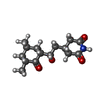


| #83: Chemical | ChemComp-3HE /  Mass: 281.347 Da / Num. of mol.: 1 / Source method: obtained synthetically / Formula: C15H23NO4 / Feature type: SUBJECT OF INVESTIGATION Mass: 281.347 Da / Num. of mol.: 1 / Source method: obtained synthetically / Formula: C15H23NO4 / Feature type: SUBJECT OF INVESTIGATION |
|---|---|
| #84: Chemical | ChemComp-SPD /  Mass: 145.246 Da / Num. of mol.: 1 / Source method: obtained synthetically / Formula: C7H19N3 Mass: 145.246 Da / Num. of mol.: 1 / Source method: obtained synthetically / Formula: C7H19N3 |
| #85: Chemical | ChemComp-MG /  Mass: 24.305 Da / Num. of mol.: 260 / Source method: obtained synthetically / Formula: Mg Mass: 24.305 Da / Num. of mol.: 260 / Source method: obtained synthetically / Formula: Mg |
-Details
| Has ligand of interest | Y |
|---|
-Experimental details
-Experiment
| Experiment | Method: ELECTRON MICROSCOPY |
|---|---|
| EM experiment | Aggregation state: PARTICLE / 3D reconstruction method: single particle reconstruction |
- Sample preparation
| Component |
| ||||||||||||||||||||||||||||||||||||||||||
|---|---|---|---|---|---|---|---|---|---|---|---|---|---|---|---|---|---|---|---|---|---|---|---|---|---|---|---|---|---|---|---|---|---|---|---|---|---|---|---|---|---|---|---|
| Source (natural) |
| ||||||||||||||||||||||||||||||||||||||||||
| Buffer solution | pH: 7.6 | ||||||||||||||||||||||||||||||||||||||||||
| Specimen | Embedding applied: NO / Shadowing applied: NO / Staining applied: NO / Vitrification applied: YES | ||||||||||||||||||||||||||||||||||||||||||
| Vitrification | Cryogen name: ETHANE |
- Electron microscopy imaging
| Experimental equipment |  Model: Titan Krios / Image courtesy: FEI Company |
|---|---|
| Microscopy | Model: FEI TITAN KRIOS |
| Electron gun | Electron source:  FIELD EMISSION GUN / Accelerating voltage: 300 kV / Illumination mode: FLOOD BEAM FIELD EMISSION GUN / Accelerating voltage: 300 kV / Illumination mode: FLOOD BEAM |
| Electron lens | Mode: BRIGHT FIELD |
| Image recording | Electron dose: 32 e/Å2 / Detector mode: SUPER-RESOLUTION / Film or detector model: GATAN K2 SUMMIT (4k x 4k) |
- Processing
| Software | Name: UCSF ChimeraX / Version: 1.1/v9 / Classification: model building / URL: https://www.rbvi.ucsf.edu/chimerax/ / Os: macOS / Type: package |
|---|---|
| CTF correction | Type: PHASE FLIPPING AND AMPLITUDE CORRECTION |
| 3D reconstruction | Resolution: 2.7 Å / Resolution method: FSC 0.143 CUT-OFF / Num. of particles: 196154 / Symmetry type: POINT |
