 Movie
Movie Controller


+ Open data

- Basic information
| Entry | Database: PDB / ID: 7r5x | |||||||||
|---|---|---|---|---|---|---|---|---|---|---|
| Title | Tankyrase 2 in complex with an inhibitor (OUL211) | |||||||||
 Components Components | (Poly [ADP-ribose] polymerase tankyrase- ...) x 2 | |||||||||
 Keywords Keywords | TRANSFERASE / Inhibitor / Complex / ADP-ribosylation / Enzyme | |||||||||
| Function / homology |  Function and homology information Function and homology informationXAV939 stabilizes AXIN / positive regulation of telomere capping / NAD+ ADP-ribosyltransferase / protein auto-ADP-ribosylation / negative regulation of telomere maintenance via telomere lengthening / protein localization to chromosome, telomeric region / NAD+-protein-aspartate ADP-ribosyltransferase activity / protein poly-ADP-ribosylation / NAD+-protein-glutamate ADP-ribosyltransferase activity / NAD+-protein mono-ADP-ribosyltransferase activity ...XAV939 stabilizes AXIN / positive regulation of telomere capping / NAD+ ADP-ribosyltransferase / protein auto-ADP-ribosylation / negative regulation of telomere maintenance via telomere lengthening / protein localization to chromosome, telomeric region / NAD+-protein-aspartate ADP-ribosyltransferase activity / protein poly-ADP-ribosylation / NAD+-protein-glutamate ADP-ribosyltransferase activity / NAD+-protein mono-ADP-ribosyltransferase activity / pericentriolar material / Transferases; Glycosyltransferases; Pentosyltransferases / NAD+ poly-ADP-ribosyltransferase activity / positive regulation of telomere maintenance via telomerase / nucleotidyltransferase activity / TCF dependent signaling in response to WNT / Degradation of AXIN / Wnt signaling pathway / Regulation of PTEN stability and activity / protein polyubiquitination / nuclear envelope / positive regulation of canonical Wnt signaling pathway / chromosome, telomeric region / Ub-specific processing proteases / Golgi membrane / perinuclear region of cytoplasm / enzyme binding / metal ion binding / nucleus / cytoplasm / cytosol Similarity search - Function | |||||||||
| Biological species |  Homo sapiens (human) Homo sapiens (human) | |||||||||
| Method |  X-RAY DIFFRACTION / SYNCHROTRON / MOLECULAR REPLACEMENT / Resolution: 2.052 Å X-RAY DIFFRACTION / SYNCHROTRON / MOLECULAR REPLACEMENT / Resolution: 2.052 Å | |||||||||
 Authors Authors | Sowa, S.T. / Lehtio, L. | |||||||||
| Funding support |  Finland, 2items Finland, 2items
| |||||||||
 Citation Citation | Journal: J.Med.Chem. / Year: 2023 Title: [1,2,4]Triazolo[3,4- b ]benzothiazole Scaffold as Versatile Nicotinamide Mimic Allowing Nanomolar Inhibition of Different PARP Enzymes. Authors: Murthy, S. / Nizi, M.G. / Maksimainen, M.M. / Massari, S. / Alaviuhkola, J. / Lippok, B.E. / Vagaggini, C. / Sowa, S.T. / Galera-Prat, A. / Ashok, Y. / Venkannagari, H. / Prunskaite- ...Authors: Murthy, S. / Nizi, M.G. / Maksimainen, M.M. / Massari, S. / Alaviuhkola, J. / Lippok, B.E. / Vagaggini, C. / Sowa, S.T. / Galera-Prat, A. / Ashok, Y. / Venkannagari, H. / Prunskaite-Hyyrylainen, R. / Dreassi, E. / Luscher, B. / Korn, P. / Tabarrini, O. / Lehtio, L. | |||||||||
| History |
|
- Structure visualization
| Structure viewer | Molecule: MolmilJmol/JSmol |
|---|
- Downloads & links
-Download
| PDBx/mmCIF format | 7r5x.cif.gz | 107.2 KB | Display | PDBx/mmCIF format |
|---|---|---|---|---|
| PDB format | pdb7r5x.ent.gz | Display | PDB format | |
| PDBx/mmJSON format | 7r5x.json.gz | Tree view | PDBx/mmJSON format | |
| Others |  Other downloads Other downloads |
-Validation report
| Arichive directory | https://data.pdbj.org/pub/pdb/validation_reports/r5/7r5xftp://data.pdbj.org/pub/pdb/validation_reports/r5/7r5x | HTTPS FTP |
|---|
-Related structure data
| Related structure data |  7r3lC  7r3oC  7r3zC 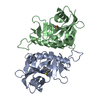 7r4aC  7r59C 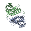 7r5dC 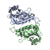 7z1vC  7z1wC  7z1yC  7z2oC 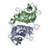 7z2qC 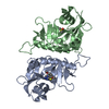 7z41C  5owsS S: Starting model for refinement C: citing same article ( |
|---|---|
| Similar structure data |
-Links
 PDBj
PDBj


- Assembly
| Deposited unit | 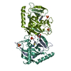
| |||||||||
|---|---|---|---|---|---|---|---|---|---|---|
| 1 | 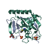
| |||||||||
| 2 | 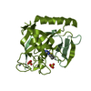
| |||||||||
| Unit cell |
| |||||||||
| Components on special symmetry positions |
|
-Components
-Poly [ADP-ribose] polymerase tankyrase- ... , 2 types, 4 molecules AAABBBAB
| #1: Protein | Mass: 19482.053 Da / Num. of mol.: 2 Source method: isolated from a genetically manipulated source Source: (gene. exp.) Homo sapiens (human) / Gene: TNKS2, PARP5B, TANK2, TNKL / Production host:  References: UniProt: Q9H2K2, NAD+ ADP-ribosyltransferase, Transferases; Glycosyltransferases; Pentosyltransferases #2: Protein/peptide | Mass: 5364.037 Da / Num. of mol.: 2 Source method: isolated from a genetically manipulated source Source: (gene. exp.) Homo sapiens (human) / Gene: TNKS2, PARP5B, TANK2, TNKL / Production host: References: UniProt: Q9H2K2, NAD+ ADP-ribosyltransferase, Transferases; Glycosyltransferases; Pentosyltransferases |
|---|
-Non-polymers , 5 types, 178 molecules 


| #3: Chemical |  Mass: 189.237 Da / Num. of mol.: 2 / Source method: obtained synthetically / Formula: C9H7N3S / Feature type: SUBJECT OF INVESTIGATION Mass: 189.237 Da / Num. of mol.: 2 / Source method: obtained synthetically / Formula: C9H7N3S / Feature type: SUBJECT OF INVESTIGATION#4: Chemical |  Mass: 65.409 Da / Num. of mol.: 2 / Source method: obtained synthetically / Formula: Zn Mass: 65.409 Da / Num. of mol.: 2 / Source method: obtained synthetically / Formula: Zn#5: Chemical | ChemComp-SO4 /  Mass: 96.063 Da / Num. of mol.: 4 / Source method: obtained synthetically / Formula: SO4 Mass: 96.063 Da / Num. of mol.: 4 / Source method: obtained synthetically / Formula: SO4#6: Chemical | ChemComp-GOL / |  Mass: 92.094 Da / Num. of mol.: 1 / Source method: obtained synthetically / Formula: C3H8O3 Mass: 92.094 Da / Num. of mol.: 1 / Source method: obtained synthetically / Formula: C3H8O3#7: Water | ChemComp-HOH / | Mass: 18.015 Da / Num. of mol.: 169 / Source method: isolated from a natural source / Formula: H2O |
|---|
-Details
| Has ligand of interest | Y |
|---|
-Experimental details
-Experiment
| Experiment | Method: X-RAY DIFFRACTION / Number of used crystals: 1 |
|---|
- Sample preparation
| Crystal | Density Matthews: 2.68 Å3/Da / Density % sol: 54.12 % |
|---|---|
| Crystal grow | Temperature: 277.15 K / Method: vapor diffusion, sitting drop Details: 100 mM Tris pH=8.5, 200mM Lithium Sulphate, 20% PEG3350 |
-Data collection
| Diffraction | Mean temperature: 100 K / Serial crystal experiment: N |
|---|---|
| Diffraction source | Source: SYNCHROTRON / Site: Diamond  / Beamline: I03 / Wavelength: 0.92023 Å / Beamline: I03 / Wavelength: 0.92023 Å |
| Detector | Type: DECTRIS EIGER2 XE 16M / Detector: PIXEL / Date: Jun 25, 2019 |
| Radiation | Protocol: SINGLE WAVELENGTH / Monochromatic (M) / Laue (L): M / Scattering type: x-ray |
| Radiation wavelength | Wavelength: 0.92023 Å / Relative weight: 1 |
| Reflection | Resolution: 2→50 Å / Num. obs: 36379 / % possible obs: 99.93 % / Redundancy: 7.9 % / CC1/2: 0.998 / Net I/σ(I): 15.2 |
| Reflection shell | Resolution: 2→2.072 Å / Num. unique obs: 3591 / CC1/2: 0.871 |
- Processing
| Software |
| ||||||||||||||||||||||||||||||||||||||||||||||||||||||||||||||||||||||||||||||||||||||||||||||||||||||||||||||||||||||||||||||||||||||||||||||||||||||
|---|---|---|---|---|---|---|---|---|---|---|---|---|---|---|---|---|---|---|---|---|---|---|---|---|---|---|---|---|---|---|---|---|---|---|---|---|---|---|---|---|---|---|---|---|---|---|---|---|---|---|---|---|---|---|---|---|---|---|---|---|---|---|---|---|---|---|---|---|---|---|---|---|---|---|---|---|---|---|---|---|---|---|---|---|---|---|---|---|---|---|---|---|---|---|---|---|---|---|---|---|---|---|---|---|---|---|---|---|---|---|---|---|---|---|---|---|---|---|---|---|---|---|---|---|---|---|---|---|---|---|---|---|---|---|---|---|---|---|---|---|---|---|---|---|---|---|---|---|---|---|---|
| Refinement | Method to determine structure: MOLECULAR REPLACEMENT Starting model: 5OWS Resolution: 2.052→48.542 Å / Cor.coef. Fo:Fc: 0.957 / Cor.coef. Fo:Fc free: 0.942 / SU B: 3.736 / SU ML: 0.104 / Cross valid method: FREE R-VALUE / ESU R: 0.164 / ESU R Free: 0.148 Details: Hydrogens have been added in their riding positions
| ||||||||||||||||||||||||||||||||||||||||||||||||||||||||||||||||||||||||||||||||||||||||||||||||||||||||||||||||||||||||||||||||||||||||||||||||||||||
| Solvent computation | Ion probe radii: 0.8 Å / Shrinkage radii: 0.8 Å / VDW probe radii: 1.2 Å / Solvent model: MASK BULK SOLVENT | ||||||||||||||||||||||||||||||||||||||||||||||||||||||||||||||||||||||||||||||||||||||||||||||||||||||||||||||||||||||||||||||||||||||||||||||||||||||
| Displacement parameters | Biso mean: 35.436 Å2
| ||||||||||||||||||||||||||||||||||||||||||||||||||||||||||||||||||||||||||||||||||||||||||||||||||||||||||||||||||||||||||||||||||||||||||||||||||||||
| Refinement step | Cycle: LAST / Resolution: 2.052→48.542 Å
| ||||||||||||||||||||||||||||||||||||||||||||||||||||||||||||||||||||||||||||||||||||||||||||||||||||||||||||||||||||||||||||||||||||||||||||||||||||||
| Refine LS restraints |
| ||||||||||||||||||||||||||||||||||||||||||||||||||||||||||||||||||||||||||||||||||||||||||||||||||||||||||||||||||||||||||||||||||||||||||||||||||||||
| LS refinement shell |
|
