 Movie
Movie Controller


+ Open data

- Basic information
| Entry | Database: PDB / ID: 7r3l | ||||||||||||
|---|---|---|---|---|---|---|---|---|---|---|---|---|---|
| Title | PARP14 catalytic domain in complex with OUL40 | ||||||||||||
 Components Components | Poly [ADP-ribose] polymerase 14 | ||||||||||||
 Keywords Keywords | TRANSFERASE / Mono-ADP-transferase / Inhibitor / Complex | ||||||||||||
| Function / homology |  Function and homology information Function and homology informationnegative regulation of tyrosine phosphorylation of STAT protein / positive regulation of interleukin-4-mediated signaling pathway / NAD+ catabolic process / Maturation of nucleoprotein / Nicotinate metabolism / positive regulation of tyrosine phosphorylation of STAT protein / Maturation of nucleoprotein / protein poly-ADP-ribosylation / NAD+-protein-glutamate ADP-ribosyltransferase activity / negative regulation of type II interferon-mediated signaling pathway ...negative regulation of tyrosine phosphorylation of STAT protein / positive regulation of interleukin-4-mediated signaling pathway / NAD+ catabolic process / Maturation of nucleoprotein / Nicotinate metabolism / positive regulation of tyrosine phosphorylation of STAT protein / Maturation of nucleoprotein / protein poly-ADP-ribosylation / NAD+-protein-glutamate ADP-ribosyltransferase activity / negative regulation of type II interferon-mediated signaling pathway / NAD+-protein mono-ADP-ribosyltransferase activity / Transferases; Glycosyltransferases; Pentosyltransferases / NAD+ poly-ADP-ribosyltransferase activity / NAD+ binding / nucleotidyltransferase activity / transcription corepressor activity / negative regulation of gene expression / innate immune response / enzyme binding / membrane / nucleus / plasma membrane / cytoplasm / cytosol Similarity search - Function | ||||||||||||
| Biological species |  Homo sapiens (human) Homo sapiens (human) | ||||||||||||
| Method |  X-RAY DIFFRACTION / SYNCHROTRON / MOLECULAR REPLACEMENT / Resolution: 2 Å X-RAY DIFFRACTION / SYNCHROTRON / MOLECULAR REPLACEMENT / Resolution: 2 Å | ||||||||||||
 Authors Authors | Maksimainen, M.M. / Murthy, S. / Lehtio, L. | ||||||||||||
| Funding support |  Finland, 3items Finland, 3items
| ||||||||||||
 Citation Citation | Journal: J.Med.Chem. / Year: 2023 Title: [1,2,4]Triazolo[3,4- b ]benzothiazole Scaffold as Versatile Nicotinamide Mimic Allowing Nanomolar Inhibition of Different PARP Enzymes. Authors: Murthy, S. / Nizi, M.G. / Maksimainen, M.M. / Massari, S. / Alaviuhkola, J. / Lippok, B.E. / Vagaggini, C. / Sowa, S.T. / Galera-Prat, A. / Ashok, Y. / Venkannagari, H. / Prunskaite- ...Authors: Murthy, S. / Nizi, M.G. / Maksimainen, M.M. / Massari, S. / Alaviuhkola, J. / Lippok, B.E. / Vagaggini, C. / Sowa, S.T. / Galera-Prat, A. / Ashok, Y. / Venkannagari, H. / Prunskaite-Hyyrylainen, R. / Dreassi, E. / Luscher, B. / Korn, P. / Tabarrini, O. / Lehtio, L. | ||||||||||||
| History |
|
- Structure visualization
| Structure viewer | Molecule: MolmilJmol/JSmol |
|---|
- Downloads & links
-Download
| PDBx/mmCIF format | 7r3l.cif.gz | 86.1 KB | Display | PDBx/mmCIF format |
|---|---|---|---|---|
| PDB format | pdb7r3l.ent.gz | 62.5 KB | Display | PDB format |
| PDBx/mmJSON format | 7r3l.json.gz | Tree view | PDBx/mmJSON format | |
| Others |  Other downloads Other downloads |
-Validation report
| Arichive directory | https://data.pdbj.org/pub/pdb/validation_reports/r3/7r3lftp://data.pdbj.org/pub/pdb/validation_reports/r3/7r3l | HTTPS FTP |
|---|
-Related structure data
| Related structure data |  7r3oC  7r3zC 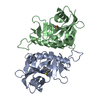 7r4aC  7r59C 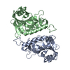 7r5dC  7r5xC 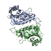 7z1vC 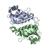 7z1wC  7z1yC  7z2oC 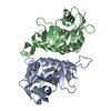 7z2qC 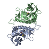 7z41C 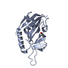 3goyS S: Starting model for refinement C: citing same article ( |
|---|---|
| Similar structure data | |
| Experimental dataset #1 | Data reference: 10.23729/0b11fe27-a545-48b0-a953-292d1e1e1d38 Data set type: diffraction image data |
-Links
 PDBj
PDBj- Assembly
| Deposited unit | 
| |||||||||
|---|---|---|---|---|---|---|---|---|---|---|
| 1 | 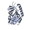
| |||||||||
| 2 | 
| |||||||||
| Unit cell |
| |||||||||
| Components on special symmetry positions |
|
-Components
| #1: Protein | Mass: 22118.590 Da / Num. of mol.: 2 Source method: isolated from a genetically manipulated source Source: (gene. exp.) Homo sapiens (human) / Gene: PARP14, BAL2, KIAA1268 / Production host:  #2: Chemical | 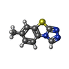  Mass: 189.237 Da / Num. of mol.: 2 / Source method: obtained synthetically / Formula: C9H7N3S / Feature type: SUBJECT OF INVESTIGATION Mass: 189.237 Da / Num. of mol.: 2 / Source method: obtained synthetically / Formula: C9H7N3S / Feature type: SUBJECT OF INVESTIGATION#3: Chemical |   Mass: 35.453 Da / Num. of mol.: 2 / Source method: obtained synthetically / Formula: Cl Mass: 35.453 Da / Num. of mol.: 2 / Source method: obtained synthetically / Formula: Cl#4: Chemical | ChemComp-PG4 / |   Mass: 194.226 Da / Num. of mol.: 1 / Source method: obtained synthetically / Formula: C8H18O5 / Comment: precipitant*YM Mass: 194.226 Da / Num. of mol.: 1 / Source method: obtained synthetically / Formula: C8H18O5 / Comment: precipitant*YM#5: Water | ChemComp-HOH / |  Mass: 18.015 Da / Num. of mol.: 141 / Source method: isolated from a natural source / Formula: H2O Mass: 18.015 Da / Num. of mol.: 141 / Source method: isolated from a natural source / Formula: H2OHas ligand of interest | Y | Has protein modification | Y | |
|---|
-Experimental details
-Experiment
| Experiment | Method: X-RAY DIFFRACTION / Number of used crystals: 1 |
|---|
- Sample preparation
| Crystal | Density Matthews: 2.79 Å3/Da / Density % sol: 55.97 % |
|---|---|
| Crystal grow | Temperature: 295 K / Method: vapor diffusion, hanging drop Details: . 0.17 M ammonium sulphate, 15% (v/v) glycerol, 27% (v/v) PEG 4000 |
-Data collection
| Diffraction | Mean temperature: 100 K / Serial crystal experiment: N |
|---|---|
| Diffraction source | Source: SYNCHROTRON / Site: ESRF  / Beamline: MASSIF-1 / Wavelength: 0.966 Å / Beamline: MASSIF-1 / Wavelength: 0.966 Å |
| Detector | Type: DECTRIS PILATUS 6M / Detector: PIXEL / Date: Oct 1, 2018 |
| Radiation | Protocol: SINGLE WAVELENGTH / Monochromatic (M) / Laue (L): M / Scattering type: x-ray |
| Radiation wavelength | Wavelength: 0.966 Å / Relative weight: 1 |
| Reflection | Resolution: 1.999→53.1 Å / Num. obs: 32725 / % possible obs: 99.2 % / Redundancy: 3.1 % / CC1/2: 0.997 / Net I/σ(I): 10.3 |
| Reflection shell | Resolution: 1.999→2.033 Å / Num. unique obs: 1587 / CC1/2: 0.842 |
- Processing
| Software |
| ||||||||||||||||||||||||||||||||||||||||||||||||||||||||||||
|---|---|---|---|---|---|---|---|---|---|---|---|---|---|---|---|---|---|---|---|---|---|---|---|---|---|---|---|---|---|---|---|---|---|---|---|---|---|---|---|---|---|---|---|---|---|---|---|---|---|---|---|---|---|---|---|---|---|---|---|---|---|
| Refinement | Method to determine structure: MOLECULAR REPLACEMENT Starting model: 3GOY Resolution: 2→53.1 Å / Cor.coef. Fo:Fc: 0.942 / Cor.coef. Fo:Fc free: 0.924 / SU B: 4.019 / SU ML: 0.109 / Cross valid method: THROUGHOUT / σ(F): 0 / ESU R: 0.158 / ESU R Free: 0.147 / Stereochemistry target values: MAXIMUM LIKELIHOOD Details: HYDROGENS HAVE BEEN ADDED IN THE RIDING POSITIONS U VALUES : REFINED INDIVIDUALLY
| ||||||||||||||||||||||||||||||||||||||||||||||||||||||||||||
| Solvent computation | Ion probe radii: 0.8 Å / Shrinkage radii: 0.8 Å / VDW probe radii: 1.2 Å / Solvent model: MASK | ||||||||||||||||||||||||||||||||||||||||||||||||||||||||||||
| Displacement parameters | Biso max: 100.9 Å2 / Biso mean: 23.308 Å2 / Biso min: 7.39 Å2
| ||||||||||||||||||||||||||||||||||||||||||||||||||||||||||||
| Refinement step | Cycle: final / Resolution: 2→53.1 Å
| ||||||||||||||||||||||||||||||||||||||||||||||||||||||||||||
| Refine LS restraints |
| ||||||||||||||||||||||||||||||||||||||||||||||||||||||||||||
| LS refinement shell | Resolution: 2→2.05 Å / Rfactor Rfree error: 0
|
