 Movie
Movie Controller

[English] 日本語
 Yorodumi
- PDB-7r5e: FtrA-P19 from Rubrivivax gelatinosus in complex with copper and m... -
+ Open data

- Basic information
| Entry | Database: PDB / ID: 7r5e | ||||||
|---|---|---|---|---|---|---|---|
| Title | FtrA-P19 from Rubrivivax gelatinosus in complex with copper and magnesium (X1) | ||||||
 Components Components | FtrA-P19 | ||||||
 Keywords Keywords | TRANSPORT PROTEIN / Copper binding / iron transport | ||||||
| Function / homology | COPPER (II) ION / DI(HYDROXYETHYL)ETHER Function and homology information Function and homology information | ||||||
| Biological species |  Rubrivivax gelatinosus (bacteria) Rubrivivax gelatinosus (bacteria) | ||||||
| Method |  X-RAY DIFFRACTION / SYNCHROTRON / MOLECULAR REPLACEMENT / molecular replacement / Resolution: 2.2 Å X-RAY DIFFRACTION / SYNCHROTRON / MOLECULAR REPLACEMENT / molecular replacement / Resolution: 2.2 Å | ||||||
 Authors Authors | Morera, S. / Vigouroux, A. | ||||||
| Funding support | 1items
| ||||||
 Citation Citation | Journal: Febs J. / Year: 2022 Title: New insights into the mechanism of iron transport through the bacterial Ftr system present in pathogens. Authors: Steunou, A.S. / Vigouroux, A. / Aumont-Nicaise, M. / Plancqueel, S. / Boussac, A. / Ouchane, S. / Morera, S. | ||||||
| History |
|
- Structure visualization
| Structure viewer | Molecule: MolmilJmol/JSmol |
|---|
- Downloads & links
-Download
| PDBx/mmCIF format | 7r5e.cif.gz | 138.8 KB | Display | PDBx/mmCIF format |
|---|---|---|---|---|
| PDB format | pdb7r5e.ent.gz | 107.3 KB | Display | PDB format |
| PDBx/mmJSON format | 7r5e.json.gz | Tree view | PDBx/mmJSON format | |
| Others |  Other downloads Other downloads |
-Validation report
| Arichive directory | https://data.pdbj.org/pub/pdb/validation_reports/r5/7r5eftp://data.pdbj.org/pub/pdb/validation_reports/r5/7r5e | HTTPS FTP |
|---|
-Related structure data
| Related structure data |  7r3pSC  7r3sC  7r4uC  7r4vC  7r4zC  7r5gC 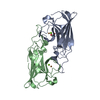 7r5pC S: Starting model for refinement C: citing same article ( |
|---|---|
| Similar structure data |
-Links
 PDBj
PDBj- Assembly
| Deposited unit | 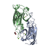
| ||||||||
|---|---|---|---|---|---|---|---|---|---|
| 1 |
| ||||||||
| Unit cell |
|
-Components
| #1: Protein | Mass: 18189.561 Da / Num. of mol.: 2 Source method: isolated from a genetically manipulated source Source: (gene. exp.) Rubrivivax gelatinosus (bacteria) / Gene: EV684_12117Production host: #2: Chemical |   Mass: 63.546 Da / Num. of mol.: 2 / Source method: obtained synthetically / Formula: Cu Mass: 63.546 Da / Num. of mol.: 2 / Source method: obtained synthetically / Formula: Cu#3: Chemical |   Mass: 24.305 Da / Num. of mol.: 3 / Source method: obtained synthetically / Formula: Mg Mass: 24.305 Da / Num. of mol.: 3 / Source method: obtained synthetically / Formula: Mg#4: Chemical | ChemComp-PEG / |   Mass: 106.120 Da / Num. of mol.: 1 / Source method: obtained synthetically / Formula: C4H10O3 Mass: 106.120 Da / Num. of mol.: 1 / Source method: obtained synthetically / Formula: C4H10O3#5: Water | ChemComp-HOH / |  Mass: 18.015 Da / Num. of mol.: 129 / Source method: isolated from a natural source / Formula: H2O Mass: 18.015 Da / Num. of mol.: 129 / Source method: isolated from a natural source / Formula: H2OHas ligand of interest | N | |
|---|
-Experimental details
-Experiment
| Experiment | Method: X-RAY DIFFRACTION / Number of used crystals: 1 |
|---|
- Sample preparation
| Crystal | Density Matthews: 2.04 Å3/Da / Density % sol: 39.58 % |
|---|---|
| Crystal grow | Temperature: 292 K / Method: vapor diffusion, hanging drop / pH: 8.5 / Details: 25% PEG 4K, 0.1 M Tris-HCl, 0.1 M MgCl2 |
-Data collection
| Diffraction | Mean temperature: 100 K / Serial crystal experiment: N |
|---|---|
| Diffraction source | Source: SYNCHROTRON / Site: SOLEIL  / Beamline: PROXIMA 2 / Wavelength: 1.73114 Å / Beamline: PROXIMA 2 / Wavelength: 1.73114 Å |
| Detector | Type: DECTRIS EIGER X 16M / Detector: PIXEL / Date: Jun 30, 2021 |
| Radiation | Protocol: SINGLE WAVELENGTH / Monochromatic (M) / Laue (L): M / Scattering type: x-ray |
| Radiation wavelength | Wavelength: 1.73114 Å / Relative weight: 1 |
| Reflection | Resolution: 2.2→27.42 Å / Num. obs: 14428 / % possible obs: 98.5 % / Redundancy: 4.1 % / Biso Wilson estimate: 27.13 Å2 / CC1/2: 0.982 / Rmerge(I) obs: 0.128 / Net I/σ(I): 8.9 |
| Reflection shell | Resolution: 2.2→2.27 Å / Rmerge(I) obs: 0.554 / Num. unique obs: 523 / CC1/2: 0.561 |
-Phasing
| Phasing | Method: molecular replacement |
|---|
- Processing
| Software |
| ||||||||||||||||||||||||||||||||||||||||||||||||||||||||||||||||||||||||||||||||||||||||||||||||||||||||||||
|---|---|---|---|---|---|---|---|---|---|---|---|---|---|---|---|---|---|---|---|---|---|---|---|---|---|---|---|---|---|---|---|---|---|---|---|---|---|---|---|---|---|---|---|---|---|---|---|---|---|---|---|---|---|---|---|---|---|---|---|---|---|---|---|---|---|---|---|---|---|---|---|---|---|---|---|---|---|---|---|---|---|---|---|---|---|---|---|---|---|---|---|---|---|---|---|---|---|---|---|---|---|---|---|---|---|---|---|---|---|
| Refinement | Method to determine structure: MOLECULAR REPLACEMENT Starting model: 7R3P Resolution: 2.2→27.42 Å / Cor.coef. Fo:Fc: 0.939 / Cor.coef. Fo:Fc free: 0.911 / SU R Cruickshank DPI: 0.318 / Cross valid method: THROUGHOUT / σ(F): 0 / SU R Blow DPI: 0.327 / SU Rfree Blow DPI: 0.205 / SU Rfree Cruickshank DPI: 0.205
| ||||||||||||||||||||||||||||||||||||||||||||||||||||||||||||||||||||||||||||||||||||||||||||||||||||||||||||
| Displacement parameters | Biso max: 99.5 Å2 / Biso mean: 23.18 Å2 / Biso min: 4.65 Å2
| ||||||||||||||||||||||||||||||||||||||||||||||||||||||||||||||||||||||||||||||||||||||||||||||||||||||||||||
| Refine analyze | Luzzati coordinate error obs: 0.24 Å | ||||||||||||||||||||||||||||||||||||||||||||||||||||||||||||||||||||||||||||||||||||||||||||||||||||||||||||
| Refinement step | Cycle: final / Resolution: 2.2→27.42 Å
| ||||||||||||||||||||||||||||||||||||||||||||||||||||||||||||||||||||||||||||||||||||||||||||||||||||||||||||
| Refine LS restraints |
| ||||||||||||||||||||||||||||||||||||||||||||||||||||||||||||||||||||||||||||||||||||||||||||||||||||||||||||
| LS refinement shell | Resolution: 2.2→2.23 Å / Rfactor Rfree error: 0 / Total num. of bins used: 35
| ||||||||||||||||||||||||||||||||||||||||||||||||||||||||||||||||||||||||||||||||||||||||||||||||||||||||||||
| Refinement TLS params. | Method: refined / Refine-ID: X-RAY DIFFRACTION
| ||||||||||||||||||||||||||||||||||||||||||||||||||||||||||||||||||||||||||||||||||||||||||||||||||||||||||||
| Refinement TLS group |
|
