 Movie
Movie Controller


+ Open data

- Basic information
| Entry | Database: PDB / ID: 7r3f | ||||||
|---|---|---|---|---|---|---|---|
| Title | Monomeric PqsE mutant E187R | ||||||
 Components Components | 2-aminobenzoylacetyl-CoA thioesterase | ||||||
 Keywords Keywords | HYDROLASE / QUORUM SENSING / PSEUDOMONAS / PROTEIN / PQS / THIOESTERASE | ||||||
| Function / homology |  Function and homology information Function and homology information2-aminobenzoylacetyl-CoA thioesterase / secondary metabolite biosynthetic process / hydrolase activity / metal ion binding Similarity search - Function | ||||||
| Biological species |  Pseudomonas aeruginosa PAO1 (bacteria) Pseudomonas aeruginosa PAO1 (bacteria) | ||||||
| Method |  X-RAY DIFFRACTION / MOLECULAR REPLACEMENT / molecular replacement / Resolution: 1.65 Å X-RAY DIFFRACTION / MOLECULAR REPLACEMENT / molecular replacement / Resolution: 1.65 Å | ||||||
 Authors Authors | Borgert, S.R. / Schmelz, S. / Blankenfeldt, W. | ||||||
| Funding support | 1items
| ||||||
 Citation Citation | Journal: Nat Commun / Year: 2022 Title: Moonlighting chaperone activity of the enzyme PqsE contributes to RhlR-controlled virulence of Pseudomonas aeruginosa. Authors: Borgert, S.R. / Henke, S. / Witzgall, F. / Schmelz, S. / Zur Lage, S. / Hotop, S.K. / Stephen, S. / Lubken, D. / Kruger, J. / Gomez, N.O. / van Ham, M. / Jansch, L. / Kalesse, M. / Pich, A. ...Authors: Borgert, S.R. / Henke, S. / Witzgall, F. / Schmelz, S. / Zur Lage, S. / Hotop, S.K. / Stephen, S. / Lubken, D. / Kruger, J. / Gomez, N.O. / van Ham, M. / Jansch, L. / Kalesse, M. / Pich, A. / Bronstrup, M. / Haussler, S. / Blankenfeldt, W. | ||||||
| History |
|
- Structure visualization
| Structure viewer | Molecule: MolmilJmol/JSmol |
|---|
- Downloads & links
-Download
| PDBx/mmCIF format | 7r3f.cif.gz | 222.7 KB | Display | PDBx/mmCIF format |
|---|---|---|---|---|
| PDB format | pdb7r3f.ent.gz | 147.5 KB | Display | PDB format |
| PDBx/mmJSON format | 7r3f.json.gz | Tree view | PDBx/mmJSON format | |
| Others |  Other downloads Other downloads |
-Validation report
| Arichive directory | https://data.pdbj.org/pub/pdb/validation_reports/r3/7r3fftp://data.pdbj.org/pub/pdb/validation_reports/r3/7r3f | HTTPS FTP |
|---|
-Related structure data
| Related structure data | 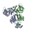 7r3eC 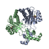 7r3gC  7r3hC  7r3iC 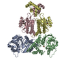 7r3jC 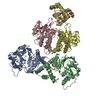 8b4aC  5hioS S: Starting model for refinement C: citing same article ( |
|---|---|
| Similar structure data |
-Links
 PDBj
PDBj


- Assembly
| Deposited unit | 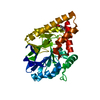
| ||||||||||
|---|---|---|---|---|---|---|---|---|---|---|---|
| 1 |
| ||||||||||
| Unit cell |
|
-Components
| #1: Protein | Mass: 34573.574 Da / Num. of mol.: 1 / Mutation: E187R Source method: isolated from a genetically manipulated source Details: Mutation at position 187: E187R / Source: (gene. exp.) Pseudomonas aeruginosa PAO1 (bacteria)Strain: ATCC 15692 / DSM 22644 / CIP 104116 / JCM 14847 / LMG 12228 / 1C / PRS 101 / PAO1 Gene: pqsE, PA1000 / Production host: References: UniProt: P20581, 2-aminobenzoylacetyl-CoA thioesterase | ||||||||
|---|---|---|---|---|---|---|---|---|---|
| #2: Chemical |   Mass: 55.845 Da / Num. of mol.: 2 / Source method: obtained synthetically / Formula: Fe Mass: 55.845 Da / Num. of mol.: 2 / Source method: obtained synthetically / Formula: Fe#3: Chemical | ChemComp-CAC / |   Mass: 136.989 Da / Num. of mol.: 1 / Source method: obtained synthetically / Formula: C2H6AsO2 Mass: 136.989 Da / Num. of mol.: 1 / Source method: obtained synthetically / Formula: C2H6AsO2#4: Chemical | ChemComp-BEZ / |   Mass: 122.121 Da / Num. of mol.: 1 / Source method: obtained synthetically / Formula: C7H6O2 Mass: 122.121 Da / Num. of mol.: 1 / Source method: obtained synthetically / Formula: C7H6O2#5: Water | ChemComp-HOH / |  Mass: 18.015 Da / Num. of mol.: 266 / Source method: isolated from a natural source / Formula: H2O Mass: 18.015 Da / Num. of mol.: 266 / Source method: isolated from a natural source / Formula: H2OHas ligand of interest | N | |
-Experimental details
-Experiment
| Experiment | Method: X-RAY DIFFRACTION / Number of used crystals: 1 |
|---|
- Sample preparation
| Crystal | Density Matthews: 2.59 Å3/Da / Density % sol: 51.64 % |
|---|---|
| Crystal grow | Temperature: 293 K / Method: vapor diffusion, sitting drop Details: 0.16 M CaAcetate, 0.08 M NaCacodylate pH 6.5, 14.4% PEG 8000, 20% Glycerol protein conc: 7.5mg/ml cryoprotectant: 10 % (v/v) (2R,3R) -(-)-2,3-Butanediol |
-Data collection
| Diffraction | Mean temperature: 100 K / Serial crystal experiment: N |
|---|---|
| Diffraction source | Source: ROTATING ANODE / Type: RIGAKU MICROMAX-007 HF / Wavelength: 1.54056 Å |
| Detector | Type: DECTRIS PILATUS3 R 300K / Detector: PIXEL / Date: Jan 25, 2021 |
| Radiation | Protocol: SINGLE WAVELENGTH / Monochromatic (M) / Laue (L): M / Scattering type: x-ray |
| Radiation wavelength | Wavelength: 1.54056 Å / Relative weight: 1 |
| Reflection | Resolution: 1.6→29.29 Å / Num. obs: 88338 / % possible obs: 99.45 % / Redundancy: 8.08 % / Biso Wilson estimate: 12.74 Å2 / Rmerge(I) obs: 0.093 / Rpim(I) all: 0.0384 / Net I/σ(I): 14.36 |
| Reflection shell | Resolution: 1.66→1.69 Å / Redundancy: 2.8 % / Rmerge(I) obs: 0.449 / Mean I/σ(I) obs: 1.11 / Num. unique obs: 8841 / Rpim(I) all: 0.3157 / % possible all: 95.72 |
-Phasing
| Phasing | Method: molecular replacement | |||||||||
|---|---|---|---|---|---|---|---|---|---|---|
| Phasing MR |
|
- Processing
| Software |
| |||||||||||||||||||||||||||||||||||||||||||||||||||||||||||||||||||||||||||||||||||||||||||||||||||||||||||||||||||||||||||||
|---|---|---|---|---|---|---|---|---|---|---|---|---|---|---|---|---|---|---|---|---|---|---|---|---|---|---|---|---|---|---|---|---|---|---|---|---|---|---|---|---|---|---|---|---|---|---|---|---|---|---|---|---|---|---|---|---|---|---|---|---|---|---|---|---|---|---|---|---|---|---|---|---|---|---|---|---|---|---|---|---|---|---|---|---|---|---|---|---|---|---|---|---|---|---|---|---|---|---|---|---|---|---|---|---|---|---|---|---|---|---|---|---|---|---|---|---|---|---|---|---|---|---|---|---|---|---|
| Refinement | Method to determine structure: MOLECULAR REPLACEMENT Starting model: 5HIO Resolution: 1.65→29.29 Å / SU ML: 0.197 / Cross valid method: FREE R-VALUE / σ(F): 1.34 / Phase error: 22.369 Stereochemistry target values: GeoStd + Monomer Library + CDL v1.2
| |||||||||||||||||||||||||||||||||||||||||||||||||||||||||||||||||||||||||||||||||||||||||||||||||||||||||||||||||||||||||||||
| Solvent computation | Shrinkage radii: 0.9 Å / VDW probe radii: 1.1 Å / Solvent model: FLAT BULK SOLVENT MODEL | |||||||||||||||||||||||||||||||||||||||||||||||||||||||||||||||||||||||||||||||||||||||||||||||||||||||||||||||||||||||||||||
| Displacement parameters | Biso mean: 16.95 Å2 | |||||||||||||||||||||||||||||||||||||||||||||||||||||||||||||||||||||||||||||||||||||||||||||||||||||||||||||||||||||||||||||
| Refinement step | Cycle: LAST / Resolution: 1.65→29.29 Å
| |||||||||||||||||||||||||||||||||||||||||||||||||||||||||||||||||||||||||||||||||||||||||||||||||||||||||||||||||||||||||||||
| Refine LS restraints |
| |||||||||||||||||||||||||||||||||||||||||||||||||||||||||||||||||||||||||||||||||||||||||||||||||||||||||||||||||||||||||||||
| LS refinement shell |
| |||||||||||||||||||||||||||||||||||||||||||||||||||||||||||||||||||||||||||||||||||||||||||||||||||||||||||||||||||||||||||||
| Refinement TLS params. | Method: refined / Refine-ID: X-RAY DIFFRACTION
| |||||||||||||||||||||||||||||||||||||||||||||||||||||||||||||||||||||||||||||||||||||||||||||||||||||||||||||||||||||||||||||
| Refinement TLS group | Refine-ID: X-RAY DIFFRACTION / Auth asym-ID: A / Label asym-ID: A
|
