 Movie
Movie Controller

[English] 日本語
 Yorodumi
- PDB-7r1n: Crystal structure of the Tetrameric C-terminal Big_2-CBM56 domain... -
+ Open data

- Basic information
| Entry | Database: PDB / ID: 7r1n | |||||||||
|---|---|---|---|---|---|---|---|---|---|---|
| Title | Crystal structure of the Tetrameric C-terminal Big_2-CBM56 domains from Paenibacillus illinoisensis (Bacillus circulans IAM1165) beta-1,3-glucanase H | |||||||||
 Components Components | Beta-1,3-glucanase bglH | |||||||||
 Keywords Keywords | HYDROLASE / laminarinase / GH16 sub-family 8 / Big_2 / CBM56 | |||||||||
| Function / homology |  Function and homology information Function and homology informationhydrolase activity, hydrolyzing O-glycosyl compounds / carbohydrate binding / carbohydrate metabolic process / metal ion binding Similarity search - Function | |||||||||
| Biological species |  Paenibacillus illinoisensis (bacteria) Paenibacillus illinoisensis (bacteria) | |||||||||
| Method |  X-RAY DIFFRACTION / SYNCHROTRON / MOLECULAR REPLACEMENT / Resolution: 2.072 Å X-RAY DIFFRACTION / SYNCHROTRON / MOLECULAR REPLACEMENT / Resolution: 2.072 Å | |||||||||
 Authors Authors | Najmudin, S. / Venditto, I. / Fontes, C.M.G.A. / Bule, P. | |||||||||
| Funding support | European Union, 2items
| |||||||||
 Citation Citation | Journal: To be published Title: Structural and biochemical characterization of C-terminal Big_2-CBM56 domains of Paenibacillus illinoisensis IAM1165 beta-1,3-glucanase H and Paenibacillus sp CBM56 Authors: Najmudin, S. / Venditto, I. / Pires, V.R. / Caseiro, C. / Correia, M.A.S. / Romao, M.J. / Carvalho, A.L. / Fontes, C.M.G.A. / Bule, P. | |||||||||
| History |
|
- Structure visualization
| Structure viewer | Molecule: MolmilJmol/JSmol |
|---|
- Downloads & links
-Download
| PDBx/mmCIF format | 7r1n.cif.gz | 474.9 KB | Display | PDBx/mmCIF format |
|---|---|---|---|---|
| PDB format | pdb7r1n.ent.gz | Display | PDB format | |
| PDBx/mmJSON format | 7r1n.json.gz | Tree view | PDBx/mmJSON format | |
| Others |  Other downloads Other downloads |
-Validation report
| Summary document | 7r1n_validation.pdf.gz | 494.3 KB | Display | wwPDB validaton report |
|---|---|---|---|---|
| Full document | 7r1n_full_validation.pdf.gz | 508.9 KB | Display | |
| Data in XML | 7r1n_validation.xml.gz | 33 KB | Display | |
| Data in CIF | 7r1n_validation.cif.gz | 45.7 KB | Display | |
| Arichive directory | https://data.pdbj.org/pub/pdb/validation_reports/r1/7r1nftp://data.pdbj.org/pub/pdb/validation_reports/r1/7r1n | HTTPS FTP |
-Related structure data
| Related structure data |  7r3tC 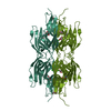 7quzS S: Starting model for refinement C: citing same article ( |
|---|---|
| Similar structure data |
-Links
 PDBj
PDBj
- Assembly
| Deposited unit | 
| ||||||||
|---|---|---|---|---|---|---|---|---|---|
| 1 |
| ||||||||
| Unit cell |
|
-Components
-Protein , 1 types, 4 molecules AAABBBCCCDDD
| #1: Protein | Mass: 21407.545 Da / Num. of mol.: 4 Source method: isolated from a genetically manipulated source Details: C-terminal Big_2-CBM56 domains / Source: (gene. exp.) Paenibacillus illinoisensis (bacteria) / Gene: bglH, beta-1,3-glucanase / Plasmid: pET28A / Production host: |
|---|
-Non-polymers , 6 types, 342 molecules 


| #2: Chemical | ChemComp-PG4 /  Mass: 194.226 Da / Num. of mol.: 4 / Source method: obtained synthetically / Formula: C8H18O5 / Comment: precipitant*YM Mass: 194.226 Da / Num. of mol.: 4 / Source method: obtained synthetically / Formula: C8H18O5 / Comment: precipitant*YM#3: Chemical | ChemComp-PEG /  Mass: 106.120 Da / Num. of mol.: 4 / Source method: obtained synthetically / Formula: C4H10O3 Mass: 106.120 Da / Num. of mol.: 4 / Source method: obtained synthetically / Formula: C4H10O3#4: Chemical |  Mass: 92.094 Da / Num. of mol.: 2 / Source method: obtained synthetically / Formula: C3H8O3 Mass: 92.094 Da / Num. of mol.: 2 / Source method: obtained synthetically / Formula: C3H8O3#5: Chemical |  Mass: 35.453 Da / Num. of mol.: 2 / Source method: obtained synthetically / Formula: Cl Mass: 35.453 Da / Num. of mol.: 2 / Source method: obtained synthetically / Formula: Cl#6: Chemical | ChemComp-MG /  Mass: 24.305 Da / Num. of mol.: 4 / Source method: obtained synthetically / Formula: Mg Mass: 24.305 Da / Num. of mol.: 4 / Source method: obtained synthetically / Formula: Mg#7: Water | ChemComp-HOH / | Mass: 18.015 Da / Num. of mol.: 326 / Source method: isolated from a natural source / Formula: H2O |
|---|
-Details
| Has ligand of interest | N |
|---|
-Experimental details
-Experiment
| Experiment | Method: X-RAY DIFFRACTION / Number of used crystals: 1 |
|---|
- Sample preparation
| Crystal | Density Matthews: 2.78 Å3/Da / Density % sol: 56 % |
|---|---|
| Crystal grow | Temperature: 292 K / Method: vapor diffusion, sitting drop / pH: 7.5 Details: 20/30/50 mg/ml Protein concentration in 0.2 Magnesium chloride hexahydrate, 0.1 M HEPES sodium salt pH 7.5. 30% v/v PEG400 |
-Data collection
| Diffraction | Mean temperature: 100 K / Serial crystal experiment: N | |||||||||||||||||||||
|---|---|---|---|---|---|---|---|---|---|---|---|---|---|---|---|---|---|---|---|---|---|---|
| Diffraction source | Source: SYNCHROTRON / Site: Diamond  / Beamline: I03 / Wavelength: 0.9795 Å / Beamline: I03 / Wavelength: 0.9795 Å | |||||||||||||||||||||
| Detector | Type: DECTRIS PILATUS 6M / Detector: PIXEL / Date: May 4, 2012 | |||||||||||||||||||||
| Radiation | Protocol: SINGLE WAVELENGTH / Monochromatic (M) / Laue (L): M / Scattering type: x-ray | |||||||||||||||||||||
| Radiation wavelength | Wavelength: 0.9795 Å / Relative weight: 1 | |||||||||||||||||||||
| Reflection | Resolution: 2.07→53.5 Å / Num. obs: 50525 / % possible obs: 98.7 % / Redundancy: 3.5 % / CC1/2: 0.989 / Rmerge(I) obs: 0.116 / Rpim(I) all: 0.097 / Rrim(I) all: 0.152 / Net I/σ(I): 6.9 | |||||||||||||||||||||
| Reflection shell | Diffraction-ID: 1
|
- Processing
| Software |
| |||||||||||||||||||||||||||||||||||||||||||||||||||||||||||||||||||||||||||||||||||||||||||||||||||||||||||||||||||||||||||||||||||||||||||||||||||||||||||
|---|---|---|---|---|---|---|---|---|---|---|---|---|---|---|---|---|---|---|---|---|---|---|---|---|---|---|---|---|---|---|---|---|---|---|---|---|---|---|---|---|---|---|---|---|---|---|---|---|---|---|---|---|---|---|---|---|---|---|---|---|---|---|---|---|---|---|---|---|---|---|---|---|---|---|---|---|---|---|---|---|---|---|---|---|---|---|---|---|---|---|---|---|---|---|---|---|---|---|---|---|---|---|---|---|---|---|---|---|---|---|---|---|---|---|---|---|---|---|---|---|---|---|---|---|---|---|---|---|---|---|---|---|---|---|---|---|---|---|---|---|---|---|---|---|---|---|---|---|---|---|---|---|---|---|---|---|
| Refinement | Method to determine structure: MOLECULAR REPLACEMENT Starting model: 7quz Resolution: 2.072→53.108 Å / Cor.coef. Fo:Fc: 0.947 / Cor.coef. Fo:Fc free: 0.899 / SU B: 13.266 / SU ML: 0.171 / Cross valid method: FREE R-VALUE / ESU R: 0.214 / ESU R Free: 0.199 Details: Hydrogens have been added in their riding positions
| |||||||||||||||||||||||||||||||||||||||||||||||||||||||||||||||||||||||||||||||||||||||||||||||||||||||||||||||||||||||||||||||||||||||||||||||||||||||||||
| Solvent computation | Ion probe radii: 0.8 Å / Shrinkage radii: 0.8 Å / VDW probe radii: 1.2 Å / Solvent model: MASK BULK SOLVENT | |||||||||||||||||||||||||||||||||||||||||||||||||||||||||||||||||||||||||||||||||||||||||||||||||||||||||||||||||||||||||||||||||||||||||||||||||||||||||||
| Displacement parameters | Biso mean: 28.204 Å2
| |||||||||||||||||||||||||||||||||||||||||||||||||||||||||||||||||||||||||||||||||||||||||||||||||||||||||||||||||||||||||||||||||||||||||||||||||||||||||||
| Refinement step | Cycle: LAST / Resolution: 2.072→53.108 Å
| |||||||||||||||||||||||||||||||||||||||||||||||||||||||||||||||||||||||||||||||||||||||||||||||||||||||||||||||||||||||||||||||||||||||||||||||||||||||||||
| Refine LS restraints |
| |||||||||||||||||||||||||||||||||||||||||||||||||||||||||||||||||||||||||||||||||||||||||||||||||||||||||||||||||||||||||||||||||||||||||||||||||||||||||||
| LS refinement shell |
| |||||||||||||||||||||||||||||||||||||||||||||||||||||||||||||||||||||||||||||||||||||||||||||||||||||||||||||||||||||||||||||||||||||||||||||||||||||||||||
| Refinement TLS params. | Method: refined / Refine-ID: X-RAY DIFFRACTION
| |||||||||||||||||||||||||||||||||||||||||||||||||||||||||||||||||||||||||||||||||||||||||||||||||||||||||||||||||||||||||||||||||||||||||||||||||||||||||||
| Refinement TLS group |
|
