 Movie
Movie Controller

[English] 日本語
 Yorodumi
- PDB-7qw6: Adenine-specific DNA methyltransferase M.BseCI complexed with Ado... -
+ Open data

- Basic information
| Entry | Database: PDB / ID: 7qw6 | ||||||
|---|---|---|---|---|---|---|---|
| Title | Adenine-specific DNA methyltransferase M.BseCI complexed with AdoHcy and cognate hemimethylated DNA duplex | ||||||
 Components Components |
| ||||||
 Keywords Keywords | TRANSFERASE / Methyltransferase / DNA-modification / Adenine-N6 methylation / Base flipping | ||||||
| Function / homology |  Function and homology information Function and homology informationsite-specific DNA-methyltransferase (adenine-specific) / site-specific DNA-methyltransferase (adenine-specific) activity / DNA restriction-modification system / methylation / DNA binding Similarity search - Function | ||||||
| Biological species |   Geobacillus stearothermophilus (bacteria) Geobacillus stearothermophilus (bacteria) | ||||||
| Method |  X-RAY DIFFRACTION / SYNCHROTRON / MOLECULAR REPLACEMENT / Resolution: 2.4 Å X-RAY DIFFRACTION / SYNCHROTRON / MOLECULAR REPLACEMENT / Resolution: 2.4 Å | ||||||
 Authors Authors | Mitsikas, D.A. / Kouyianou, K. / Kotsifaki, D. / Providaki, M. / Bouriotis, V. / Glykos, N.M. / Kokkinidis, M. | ||||||
| Funding support |  Greece, 1items Greece, 1items
| ||||||
 Citation Citation | Journal: To Be Published Title: Structure of M.BseCI DNA methyltransferase from Geobacillus stearothermophilus. Authors: Mitsikas, D.A. / Kouyianou, K. / Kotsifaki, D. / Providaki, M. / Bouriotis, V. / Glykos, N.M. / Kokkinidis, M. | ||||||
| History |
|
- Structure visualization
| Structure viewer | Molecule: MolmilJmol/JSmol |
|---|
- Downloads & links
-Download
| PDBx/mmCIF format | 7qw6.cif.gz | 299.5 KB | Display | PDBx/mmCIF format |
|---|---|---|---|---|
| PDB format | pdb7qw6.ent.gz | 210.6 KB | Display | PDB format |
| PDBx/mmJSON format | 7qw6.json.gz | Tree view | PDBx/mmJSON format | |
| Others |  Other downloads Other downloads |
-Validation report
| Arichive directory | https://data.pdbj.org/pub/pdb/validation_reports/qw/7qw6ftp://data.pdbj.org/pub/pdb/validation_reports/qw/7qw6 | HTTPS FTP |
|---|
-Related structure data
| Related structure data | 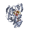 7qw5SC 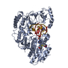 7qw7C 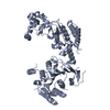 7qw8C S: Starting model for refinement C: citing same article ( |
|---|---|
| Similar structure data |
-Links
 PDBj
PDBj- Assembly
| Deposited unit | 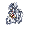
| ||||||||||||
|---|---|---|---|---|---|---|---|---|---|---|---|---|---|
| 1 |
| ||||||||||||
| Unit cell |
|
-Components
| #1: Protein | Mass: 67761.094 Da / Num. of mol.: 1 Source method: isolated from a genetically manipulated source Source: (gene. exp.) Geobacillus stearothermophilus (bacteria)Gene: bseCIM / Production host: References: UniProt: P43423, site-specific DNA-methyltransferase (adenine-specific) |
|---|---|
| #2: DNA chain | Mass: 3045.004 Da / Num. of mol.: 1 / Source method: obtained synthetically / Source: (synth.) Geobacillus stearothermophilus (bacteria) |
| #3: DNA chain | Mass: 3059.031 Da / Num. of mol.: 1 / Source method: obtained synthetically / Source: (synth.) Geobacillus stearothermophilus (bacteria) |
| #4: Chemical | ChemComp-SAH /   Mass: 384.411 Da / Num. of mol.: 1 / Source method: obtained synthetically / Formula: C14H20N6O5S / Feature type: SUBJECT OF INVESTIGATION Mass: 384.411 Da / Num. of mol.: 1 / Source method: obtained synthetically / Formula: C14H20N6O5S / Feature type: SUBJECT OF INVESTIGATION |
| #5: Water | ChemComp-HOH /  Mass: 18.015 Da / Num. of mol.: 187 / Source method: isolated from a natural source / Formula: H2O Mass: 18.015 Da / Num. of mol.: 187 / Source method: isolated from a natural source / Formula: H2O |
| Has ligand of interest | Y |
| Has protein modification | N |
-Experimental details
-Experiment
| Experiment | Method: X-RAY DIFFRACTION / Number of used crystals: 1 |
|---|
- Sample preparation
| Crystal | Density Matthews: 2.46 Å3/Da / Density % sol: 50.05 % |
|---|---|
| Crystal grow | Temperature: 293 K / Method: vapor diffusion, hanging drop / pH: 9.5 / Details: Glycerol ethoxylate, Tris-HCl, paraffin oil |
-Data collection
| Diffraction | Mean temperature: 100 K / Serial crystal experiment: N |
|---|---|
| Diffraction source | Source: SYNCHROTRON / Site: ALBA  / Beamline: XALOC / Wavelength: 0.97923 Å / Beamline: XALOC / Wavelength: 0.97923 Å |
| Detector | Type: DECTRIS PILATUS 6M / Detector: PIXEL / Date: Feb 7, 2015 |
| Radiation | Protocol: SINGLE WAVELENGTH / Monochromatic (M) / Laue (L): M / Scattering type: x-ray |
| Radiation wavelength | Wavelength: 0.97923 Å / Relative weight: 1 |
| Reflection | Resolution: 2.4→52.03 Å / Num. obs: 24491 / % possible obs: 92.4 % / Redundancy: 5.3 % / Biso Wilson estimate: 54.29 Å2 / CC1/2: 0.995 / Rmerge(I) obs: 0.11 / Rpim(I) all: 0.048 / Rrim(I) all: 0.12 / Χ2: 1.03 / Net I/σ(I): 7.8 |
| Reflection shell | Resolution: 2.4→2.49 Å / Redundancy: 5.3 % / Rmerge(I) obs: 0.999 / Mean I/σ(I) obs: 1.4 / Num. unique obs: 2662 / Rpim(I) all: 0.431 / % possible all: 94.9 |
- Processing
| Software |
| ||||||||||||||||||||||||||||||||||||||||||||||||||||||||||||||||||||||||||||||||||||||||||||||||||||
|---|---|---|---|---|---|---|---|---|---|---|---|---|---|---|---|---|---|---|---|---|---|---|---|---|---|---|---|---|---|---|---|---|---|---|---|---|---|---|---|---|---|---|---|---|---|---|---|---|---|---|---|---|---|---|---|---|---|---|---|---|---|---|---|---|---|---|---|---|---|---|---|---|---|---|---|---|---|---|---|---|---|---|---|---|---|---|---|---|---|---|---|---|---|---|---|---|---|---|---|---|---|
| Refinement | Method to determine structure: MOLECULAR REPLACEMENT Starting model: 7QW5 Resolution: 2.4→42.93 Å / SU ML: 0.2904 / Cross valid method: FREE R-VALUE / σ(F): 1.35 / Phase error: 26.0864 Stereochemistry target values: GeoStd + Monomer Library + CDL v1.2
| ||||||||||||||||||||||||||||||||||||||||||||||||||||||||||||||||||||||||||||||||||||||||||||||||||||
| Solvent computation | Shrinkage radii: 0.9 Å / VDW probe radii: 1.11 Å / Solvent model: FLAT BULK SOLVENT MODEL / Bsol: 46.2 Å2 / ksol: 0.3 e/Å3 | ||||||||||||||||||||||||||||||||||||||||||||||||||||||||||||||||||||||||||||||||||||||||||||||||||||
| Displacement parameters | Biso mean: 62.94 Å2 | ||||||||||||||||||||||||||||||||||||||||||||||||||||||||||||||||||||||||||||||||||||||||||||||||||||
| Refinement step | Cycle: LAST / Resolution: 2.4→42.93 Å
| ||||||||||||||||||||||||||||||||||||||||||||||||||||||||||||||||||||||||||||||||||||||||||||||||||||
| Refine LS restraints |
| ||||||||||||||||||||||||||||||||||||||||||||||||||||||||||||||||||||||||||||||||||||||||||||||||||||
| LS refinement shell |
| ||||||||||||||||||||||||||||||||||||||||||||||||||||||||||||||||||||||||||||||||||||||||||||||||||||
| Refinement TLS params. | Method: refined / Refine-ID: X-RAY DIFFRACTION
| ||||||||||||||||||||||||||||||||||||||||||||||||||||||||||||||||||||||||||||||||||||||||||||||||||||
| Refinement TLS group | Refine-ID: X-RAY DIFFRACTION
|
