 Movie
Movie Controller


+ Open data

- Basic information
| Entry | Database: PDB / ID: 7qr4 | ||||||||||||
|---|---|---|---|---|---|---|---|---|---|---|---|---|---|
| Title | human CPEB3 HDV-like ribozyme | ||||||||||||
 Components Components |
| ||||||||||||
 Keywords Keywords | RNA / Ribozyme HDV-like CPEB3 intron brain | ||||||||||||
| Function / homology |  Function and homology information Function and homology information | ||||||||||||
| Biological species |  Homo sapiens (human) Homo sapiens (human) | ||||||||||||
| Method |  X-RAY DIFFRACTION / SYNCHROTRON / MOLECULAR REPLACEMENT / Resolution: 2.83 Å X-RAY DIFFRACTION / SYNCHROTRON / MOLECULAR REPLACEMENT / Resolution: 2.83 Å | ||||||||||||
 Authors Authors | Przytula-Mally, A.I. / Engilberge, S. / Johannsen, S. / Olieric, V. / Masquida, B. / Sigel, R.K.O. | ||||||||||||
| Funding support |  Switzerland, 3items Switzerland, 3items
| ||||||||||||
 Citation Citation | Journal: Biorxiv / Year: 2022 Title: Anticodon-like loop-mediated dimerization in the crystal structures of HdV-like CPEB3 ribozymes Authors: Przytula-Mally, A.I. / Engilberge, S. / Johannsen, S. / Olieric, V. / Masquida, B. / Sigel, R.K. | ||||||||||||
| History |
|
- Structure visualization
| Structure viewer | Molecule: MolmilJmol/JSmol |
|---|
- Downloads & links
-Download
| PDBx/mmCIF format | 7qr4.cif.gz | 149.2 KB | Display | PDBx/mmCIF format |
|---|---|---|---|---|
| PDB format | pdb7qr4.ent.gz | 96.1 KB | Display | PDB format |
| PDBx/mmJSON format | 7qr4.json.gz | Tree view | PDBx/mmJSON format | |
| Others |  Other downloads Other downloads |
-Validation report
| Arichive directory | https://data.pdbj.org/pub/pdb/validation_reports/qr/7qr4ftp://data.pdbj.org/pub/pdb/validation_reports/qr/7qr4 | HTTPS FTP |
|---|
-Related structure data
| Related structure data |  7qr3C 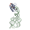 1drzS S: Starting model for refinement C: citing same article ( |
|---|---|
| Similar structure data |
-Links
 PDBj
PDBj


- Assembly
| Deposited unit | 
| ||||||||||||
|---|---|---|---|---|---|---|---|---|---|---|---|---|---|
| 1 | 
| ||||||||||||
| Unit cell |
|
-Components
| #1: Protein | Mass: 10582.399 Da / Num. of mol.: 1 Source method: isolated from a genetically manipulated source Source: (gene. exp.) Homo sapiens (human) / Gene: SNRPA / Plasmid: p11U1ADb / Production host: Expression vector pET-mod (others) / References: UniProt: M0R221 |
|---|---|
| #2: RNA chain | Mass: 22114.150 Da / Num. of mol.: 1 / Source method: obtained synthetically / Source: (synth.) Homo sapiens (human) / References: GenBank: 21540012 |
| #3: Water | ChemComp-HOH /  Mass: 18.015 Da / Num. of mol.: 3 / Source method: isolated from a natural source / Formula: H2O Mass: 18.015 Da / Num. of mol.: 3 / Source method: isolated from a natural source / Formula: H2O |
-Experimental details
-Experiment
| Experiment | Method: X-RAY DIFFRACTION / Number of used crystals: 1 |
|---|
- Sample preparation
| Crystal | Density Matthews: 3.62 Å3/Da / Density % sol: 66.05 % |
|---|---|
| Crystal grow | Temperature: 277 K / Method: vapor diffusion Details: 20-24% PEG 3350, 0.2 M sodium citrate tribasic dihydrate and 30-200 mM HEPES-KOH pH 7.5 |
-Data collection
| Diffraction | Mean temperature: 110 K / Serial crystal experiment: N |
|---|---|
| Diffraction source | Source: SYNCHROTRON / Site: SLS / Beamline: X06DA / Wavelength: 1.00004 Å |
| Detector | Type: DECTRIS PILATUS 2M-F / Detector: PIXEL / Date: Jul 5, 2018 |
| Radiation | Protocol: SINGLE WAVELENGTH / Monochromatic (M) / Laue (L): M / Scattering type: x-ray |
| Radiation wavelength | Wavelength: 1.00004 Å / Relative weight: 1 |
| Reflection | Resolution: 2.825→68.055 Å / Num. obs: 6828 / % possible obs: 92.6 % / Redundancy: 12.8 % / Biso Wilson estimate: 93.31 Å2 / CC1/2: 0.998 / Net I/σ(I): 16.7 |
| Reflection shell | Resolution: 2.825→3.124 Å / Num. unique obs: 342 / CC1/2: 0.574 |
- Processing
| Software |
| ||||||||||||||||||||||||||||||||||||||||
|---|---|---|---|---|---|---|---|---|---|---|---|---|---|---|---|---|---|---|---|---|---|---|---|---|---|---|---|---|---|---|---|---|---|---|---|---|---|---|---|---|---|
| Refinement | Method to determine structure: MOLECULAR REPLACEMENT Starting model: 1drz Resolution: 2.83→65.94 Å / SU ML: 0.389 / Cross valid method: FREE R-VALUE / σ(F): 1.36 / Phase error: 36.1353 Stereochemistry target values: GeoStd + Monomer Library + CDL v1.2
| ||||||||||||||||||||||||||||||||||||||||
| Solvent computation | Shrinkage radii: 0.9 Å / VDW probe radii: 1.11 Å / Solvent model: FLAT BULK SOLVENT MODEL | ||||||||||||||||||||||||||||||||||||||||
| Displacement parameters | Biso mean: 95.87 Å2 | ||||||||||||||||||||||||||||||||||||||||
| Refinement step | Cycle: LAST / Resolution: 2.83→65.94 Å
| ||||||||||||||||||||||||||||||||||||||||
| Refine LS restraints |
| ||||||||||||||||||||||||||||||||||||||||
| LS refinement shell |
| ||||||||||||||||||||||||||||||||||||||||
| Refinement TLS params. | Method: refined / Origin x: 0.0485657081387 Å / Origin y: 3.17551941062 Å / Origin z: 8.32629500816 Å
| ||||||||||||||||||||||||||||||||||||||||
| Refinement TLS group | Selection details: all |
