 Movie
Movie Controller

[English] 日本語
 Yorodumi
- PDB-7qf7: Orthorhombic crystal structure of PTG CBM21 in complex with beta-... -
+ Open data

- Basic information
| Entry | Database: PDB / ID: 7qf7 | ||||||
|---|---|---|---|---|---|---|---|
| Title | Orthorhombic crystal structure of PTG CBM21 in complex with beta-cyclodextrin | ||||||
 Components Components | Protein phosphatase 1 regulatory subunit 3C | ||||||
 Keywords Keywords | SUGAR BINDING PROTEIN / carbohydrate binding / immunoglobulin-like fold | ||||||
| Function / homology |  Function and homology information Function and homology informationglycogen binding / regulation of glycogen biosynthetic process / protein phosphatase type 1 complex / protein phosphatase 1 binding / glycogen biosynthetic process / glycogen metabolic process / protein serine/threonine phosphatase activity / Myoclonic epilepsy of Lafora / Glycogen synthesis / protein phosphatase binding ...glycogen binding / regulation of glycogen biosynthetic process / protein phosphatase type 1 complex / protein phosphatase 1 binding / glycogen biosynthetic process / glycogen metabolic process / protein serine/threonine phosphatase activity / Myoclonic epilepsy of Lafora / Glycogen synthesis / protein phosphatase binding / protein-macromolecule adaptor activity / cytosol Similarity search - Function | ||||||
| Biological species |  Homo sapiens (human) Homo sapiens (human) | ||||||
| Method |  X-RAY DIFFRACTION / SYNCHROTRON / MOLECULAR REPLACEMENT / molecular replacement / Resolution: 1.47 Å X-RAY DIFFRACTION / SYNCHROTRON / MOLECULAR REPLACEMENT / molecular replacement / Resolution: 1.47 Å | ||||||
 Authors Authors | Semrau, M.S. / Storici, P. / Lolli, G. | ||||||
| Funding support | 1items
| ||||||
 Citation Citation | Journal: Nat Commun / Year: 2022 Title: Molecular architecture of the glycogen- committed PP1/PTG holoenzyme. Authors: Semrau, M.S. / Giachin, G. / Covaceuszach, S. / Cassetta, A. / Demitri, N. / Storici, P. / Lolli, G. | ||||||
| History |
|
- Structure visualization
| Structure viewer | Molecule: MolmilJmol/JSmol |
|---|
- Downloads & links
-Download
| PDBx/mmCIF format | 7qf7.cif.gz | 76.8 KB | Display | PDBx/mmCIF format |
|---|---|---|---|---|
| PDB format | pdb7qf7.ent.gz | 54.9 KB | Display | PDB format |
| PDBx/mmJSON format | 7qf7.json.gz | Tree view | PDBx/mmJSON format | |
| Others |  Other downloads Other downloads |
-Validation report
| Arichive directory | https://data.pdbj.org/pub/pdb/validation_reports/qf/7qf7ftp://data.pdbj.org/pub/pdb/validation_reports/qf/7qf7 | HTTPS FTP |
|---|
-Related structure data
| Related structure data | 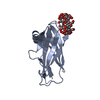 7qfaC  7qfbC  7qm2C  2eefS S: Starting model for refinement C: citing same article ( |
|---|---|
| Similar structure data |
-Links
 PDBj
PDBj


- Assembly
| Deposited unit | 
| ||||||||
|---|---|---|---|---|---|---|---|---|---|
| 1 |
| ||||||||
| Unit cell |
|
-Components
| #1: Protein | Mass: 15647.529 Da / Num. of mol.: 1 / Fragment: CBM21 domain (residues 132-264) / Mutation: First residue S derives from the expression tag Source method: isolated from a genetically manipulated source Source: (gene. exp.) Homo sapiens (human) / Gene: PPP1R3C, PPP1R5 / Production host:  |
|---|---|
| #2: Polysaccharide | Cycloheptakis-(1-4)-(alpha-D-glucopyranose)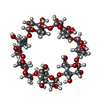  Type: oligosaccharide, Oligosaccharide / Class: Drug delivery / Mass: 1153.001 Da / Num. of mol.: 1 Type: oligosaccharide, Oligosaccharide / Class: Drug delivery / Mass: 1153.001 Da / Num. of mol.: 1Source method: isolated from a genetically manipulated source Details: cyclic oligosaccharide / References: beta-cyclodextrin |
| #3: Chemical | ChemComp-NA /   Mass: 22.990 Da / Num. of mol.: 1 / Source method: obtained synthetically / Formula: Na Mass: 22.990 Da / Num. of mol.: 1 / Source method: obtained synthetically / Formula: Na |
| #4: Water | ChemComp-HOH /  Mass: 18.015 Da / Num. of mol.: 134 / Source method: isolated from a natural source / Formula: H2O Mass: 18.015 Da / Num. of mol.: 134 / Source method: isolated from a natural source / Formula: H2O |
| Has ligand of interest | Y |
-Experimental details
-Experiment
| Experiment | Method: X-RAY DIFFRACTION / Number of used crystals: 1 |
|---|
- Sample preparation
| Crystal | Density Matthews: 2.46 Å3/Da / Density % sol: 49.91 % / Mosaicity: 0.16 ° |
|---|---|
| Crystal grow | Temperature: 293 K / Method: vapor diffusion, sitting drop / pH: 5.5 / Details: 3 M NaCl |
-Data collection
| Diffraction | Mean temperature: 100 K / Serial crystal experiment: N | ||||||||||||||||||||||||||||||
|---|---|---|---|---|---|---|---|---|---|---|---|---|---|---|---|---|---|---|---|---|---|---|---|---|---|---|---|---|---|---|---|
| Diffraction source | Source: SYNCHROTRON / Site: ELETTRA  / Beamline: 11.2C / Wavelength: 0.9718 Å / Beamline: 11.2C / Wavelength: 0.9718 Å | ||||||||||||||||||||||||||||||
| Detector | Type: DECTRIS PILATUS 6M / Detector: PIXEL / Date: Dec 15, 2020 | ||||||||||||||||||||||||||||||
| Radiation | Protocol: SINGLE WAVELENGTH / Monochromatic (M) / Laue (L): M / Scattering type: x-ray | ||||||||||||||||||||||||||||||
| Radiation wavelength | Wavelength: 0.9718 Å / Relative weight: 1 | ||||||||||||||||||||||||||||||
| Reflection | Resolution: 1.47→44.074 Å / Num. obs: 26941 / % possible obs: 99.9 % / Redundancy: 6.8 % / CC1/2: 0.999 / Rmerge(I) obs: 0.047 / Rpim(I) all: 0.02 / Rrim(I) all: 0.052 / Net I/σ(I): 17.1 | ||||||||||||||||||||||||||||||
| Reflection shell | Diffraction-ID: 1
|
-Phasing
| Phasing | Method: molecular replacement |
|---|
- Processing
| Software |
| ||||||||||||||||||||||||||||||||||||||||||||||||||
|---|---|---|---|---|---|---|---|---|---|---|---|---|---|---|---|---|---|---|---|---|---|---|---|---|---|---|---|---|---|---|---|---|---|---|---|---|---|---|---|---|---|---|---|---|---|---|---|---|---|---|---|
| Refinement | Method to determine structure: MOLECULAR REPLACEMENT Starting model: 2EEF Resolution: 1.47→44.074 Å / SU ML: 0.14 / Cross valid method: FREE R-VALUE / σ(F): 1.34 / Phase error: 21.96 / Stereochemistry target values: ML
| ||||||||||||||||||||||||||||||||||||||||||||||||||
| Solvent computation | Shrinkage radii: 0.9 Å / VDW probe radii: 1.11 Å / Solvent model: FLAT BULK SOLVENT MODEL | ||||||||||||||||||||||||||||||||||||||||||||||||||
| Displacement parameters | Biso max: 86.34 Å2 / Biso mean: 33.8912 Å2 / Biso min: 17.14 Å2 | ||||||||||||||||||||||||||||||||||||||||||||||||||
| Refinement step | Cycle: final / Resolution: 1.47→44.074 Å
| ||||||||||||||||||||||||||||||||||||||||||||||||||
| Refine LS restraints |
| ||||||||||||||||||||||||||||||||||||||||||||||||||
| LS refinement shell | Refine-ID: X-RAY DIFFRACTION / Rfactor Rfree error: 0 / % reflection obs: 100 %
|
