| 登録情報 | データベース: PDB / ID: 7q6g
|
|---|
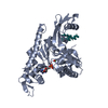
| タイトル | Xenorhabdus poinarii FtsA 1-396 ADP |
|---|
 要素 要素 | Cell division protein FtsA |
|---|
 キーワード キーワード | CELL CYCLE / Bacterial cell division / Divisome / Actin homologue |
|---|
| 機能・相同性 |  機能・相同性情報 機能・相同性情報
FtsZ-dependent cytokinesis / cell division site / cytoplasmic side of plasma membrane / nucleotide binding / metal ion binding類似検索 - 分子機能 Dna Ligase; domain 1 - #110 / Cell division protein FtsA / SHS2 domain inserted in FTSA / Cell division protein FtsA / SHS2 domain inserted in FtsA / Cell division protein FtsA / : / Dna Ligase; domain 1 / ATPase, nucleotide binding domain / 2-Layer Sandwich / Alpha Beta類似検索 - ドメイン・相同性 ADENOSINE-5'-DIPHOSPHATE / Cell division protein FtsA類似検索 - 構成要素 |
|---|
| 生物種 |  Xenorhabdus poinarii G6 (バクテリア) Xenorhabdus poinarii G6 (バクテリア) |
|---|
| 手法 |  X線回折 / シンクロトロン / 分子置換 / 解像度: 2.8 Å X線回折 / シンクロトロン / 分子置換 / 解像度: 2.8 Å |
|---|
 データ登録者 データ登録者 | Nierhaus, T. / Kureisaite-Ciziene, D. / Lowe, J. |
|---|
| 資金援助 |  英国, 2件 英国, 2件 | 組織 | 認可番号 | 国 |
|---|
| Wellcome Trust | 202754/Z/16/Z | 英国 | | Medical Research Council (MRC, United Kingdom) | U105184326 | 英国 |
|
|---|
 引用 引用 | ジャーナル: Nat Microbiol / 年: 2022
タイトル: Bacterial divisome protein FtsA forms curved antiparallel double filaments when binding to FtsN.
著者: Nierhaus, T. / McLaughlin, S.H. / Burmann, F. / Kureisaite-Ciziene, D. / Maslen, S.L. / Skehel, J.M. / Yu, C.W.H. / Freund, S.M.V. / Funke, L.F.H. / Chin, J.W. / Lowe, J. |
|---|
| 履歴 | | 登録 | 2021年11月7日 | 登録サイト: PDBE / 処理サイト: PDBE |
|---|
| 改定 1.0 | 2022年9月21日 | Provider: repository / タイプ: Initial release |
|---|
| 改定 1.1 | 2022年9月28日 | Group: Database references / カテゴリ: citation / citation_author
Item: _citation.country / _citation.journal_abbrev ..._citation.country / _citation.journal_abbrev / _citation.journal_id_ISSN / _citation.pdbx_database_id_DOI / _citation.title / _citation.year / _citation_author.name |
|---|
| 改定 1.2 | 2022年10月5日 | Group: Database references / カテゴリ: citation / Item: _citation.pdbx_database_id_PubMed / _citation.title |
|---|
| 改定 1.3 | 2022年10月12日 | Group: Database references / カテゴリ: citation
Item: _citation.journal_volume / _citation.page_first / _citation.page_last |
|---|
| 改定 1.4 | 2024年1月31日 | Group: Data collection / Refinement description
カテゴリ: chem_comp_atom / chem_comp_bond / pdbx_initial_refinement_model |
|---|
|
|---|
 ムービー
ムービー コントローラー


 データを開く
 基本情報
基本情報 構造の表示
構造の表示 ダウンロードとリンク
 その他のダウンロード

 PDBj
PDBj
 集合体
集合体

 分子量: 427.201 Da / 分子数: 1 / 由来タイプ: 合成 / 式: C10H15N5O10P2 / タイプ: SUBJECT OF INVESTIGATION / コメント: ADP, エネルギー貯蔵分子*YM
 分子量: 24.305 Da / 分子数: 1 / 由来タイプ: 合成 / 式: Mg
 試料調製
試料調製 解析
解析