 Movie
Movie Controller

[English] 日本語
 Yorodumi
- PDB-7q3t: Crystal structure of the OmpK36 D insertion chimera from Klebsiel... -
+ Open data

- Basic information
| Entry | Database: PDB / ID: 7q3t | ||||||
|---|---|---|---|---|---|---|---|
| Title | Crystal structure of the OmpK36 D insertion chimera from Klebsiella pneumonia | ||||||
 Components Components | OmpK36 | ||||||
 Keywords Keywords | MEMBRANE PROTEIN / outer membrane porin | ||||||
| Function / homology |  Function and homology information Function and homology informationporin activity / pore complex / cell outer membrane / monoatomic ion transmembrane transport / metal ion binding Similarity search - Function | ||||||
| Biological species |  Klebsiella pneumoniae (bacteria) Klebsiella pneumoniae (bacteria) | ||||||
| Method |  X-RAY DIFFRACTION / SYNCHROTRON / MOLECULAR REPLACEMENT / Resolution: 1.788 Å X-RAY DIFFRACTION / SYNCHROTRON / MOLECULAR REPLACEMENT / Resolution: 1.788 Å | ||||||
 Authors Authors | Kwong, H. / Beis, K. | ||||||
| Funding support |  United Kingdom, 1items United Kingdom, 1items
| ||||||
 Citation Citation | Journal: Plos Pathog. / Year: 2022 Title: Widespread emergence of OmpK36 loop 3 insertions among multidrug-resistant clones of Klebsiella pneumoniae. Authors: David, S. / Wong, J.L.C. / Sanchez-Garrido, J. / Kwong, H.S. / Low, W.W. / Morecchiato, F. / Giani, T. / Rossolini, G.M. / Brett, S.J. / Clements, A. / Beis, K. / Aanensen, D.M. / Frankel, G. | ||||||
| History |
|
- Structure visualization
| Structure viewer | Molecule: MolmilJmol/JSmol |
|---|
- Downloads & links
-Download
| PDBx/mmCIF format | 7q3t.cif.gz | 436 KB | Display | PDBx/mmCIF format |
|---|---|---|---|---|
| PDB format | pdb7q3t.ent.gz | 355.4 KB | Display | PDB format |
| PDBx/mmJSON format | 7q3t.json.gz | Tree view | PDBx/mmJSON format | |
| Others |  Other downloads Other downloads |
-Validation report
| Arichive directory | https://data.pdbj.org/pub/pdb/validation_reports/q3/7q3tftp://data.pdbj.org/pub/pdb/validation_reports/q3/7q3t | HTTPS FTP |
|---|
-Related structure data
| Related structure data |  7pzfC  6rd3S S: Starting model for refinement C: citing same article ( |
|---|---|
| Similar structure data |
-Links
 PDBj
PDBj
- Assembly
| Deposited unit | 
| ||||||||
|---|---|---|---|---|---|---|---|---|---|
| 1 | 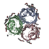
| ||||||||
| 2 | 
| ||||||||
| Unit cell |
|
-Components
| #1: Protein | Mass: 38174.012 Da / Num. of mol.: 6 / Mutation: D115 insertion Source method: isolated from a genetically manipulated source Source: (gene. exp.) Klebsiella pneumoniae (bacteria) / Gene: ompK36 / Production host: #2: Chemical | ChemComp-C8E / (   Mass: 306.438 Da / Num. of mol.: 47 / Source method: obtained synthetically / Formula: C16H34O5 / Comment: C8E, detergent*YM Mass: 306.438 Da / Num. of mol.: 47 / Source method: obtained synthetically / Formula: C16H34O5 / Comment: C8E, detergent*YM#3: Chemical | ChemComp-LI /   Mass: 6.941 Da / Num. of mol.: 12 / Source method: obtained synthetically / Formula: Li Mass: 6.941 Da / Num. of mol.: 12 / Source method: obtained synthetically / Formula: Li#4: Water | ChemComp-HOH / |  Mass: 18.015 Da / Num. of mol.: 941 / Source method: isolated from a natural source / Formula: H2O Mass: 18.015 Da / Num. of mol.: 941 / Source method: isolated from a natural source / Formula: H2OHas ligand of interest | N | |
|---|
-Experimental details
-Experiment
| Experiment | Method: X-RAY DIFFRACTION / Number of used crystals: 1 |
|---|
- Sample preparation
| Crystal | Density Matthews: 2.71 Å3/Da / Density % sol: 54.56 % |
|---|---|
| Crystal grow | Temperature: 293 K / Method: vapor diffusion, hanging drop / pH: 5.6 Details: 0.1 M Lithium sulfate, 0.1 M Sodium citrate, and 10% PEG 4000 (pH 5.6) |
-Data collection
| Diffraction | Mean temperature: 100 K / Serial crystal experiment: N |
|---|---|
| Diffraction source | Source: SYNCHROTRON / Site: Diamond / Beamline: I24 / Wavelength: 0.9686 Å |
| Detector | Type: DECTRIS PILATUS 6M / Detector: PIXEL / Date: Feb 21, 2020 |
| Radiation | Protocol: SINGLE WAVELENGTH / Monochromatic (M) / Laue (L): M / Scattering type: x-ray |
| Radiation wavelength | Wavelength: 0.9686 Å / Relative weight: 1 |
| Reflection | Resolution: 1.78→78.6 Å / Num. obs: 221416 / % possible obs: 97.4 % / Redundancy: 3.5 % / Biso Wilson estimate: 22.84 Å2 / CC1/2: 0.99 / Rmerge(I) obs: 0.154 / Net I/σ(I): 6 |
| Reflection shell | Resolution: 1.78→1.81 Å / Rmerge(I) obs: 1.296 / Mean I/σ(I) obs: 1 / Num. unique obs: 10907 / CC1/2: 0.34 / % possible all: 96.2 |
- Processing
| Software |
| ||||||||||||||||||||||||||||||||||||||||||||||||||||||||||||||||||||||||||||||||||||||||||||||||||||||||||||||||||
|---|---|---|---|---|---|---|---|---|---|---|---|---|---|---|---|---|---|---|---|---|---|---|---|---|---|---|---|---|---|---|---|---|---|---|---|---|---|---|---|---|---|---|---|---|---|---|---|---|---|---|---|---|---|---|---|---|---|---|---|---|---|---|---|---|---|---|---|---|---|---|---|---|---|---|---|---|---|---|---|---|---|---|---|---|---|---|---|---|---|---|---|---|---|---|---|---|---|---|---|---|---|---|---|---|---|---|---|---|---|---|---|---|---|---|---|
| Refinement | Method to determine structure: MOLECULAR REPLACEMENT Starting model: 6rd3 Resolution: 1.788→78.6 Å / Cor.coef. Fo:Fc: 0.94 / Cor.coef. Fo:Fc free: 0.925 / SU R Cruickshank DPI: 0.115 / Cross valid method: THROUGHOUT / σ(F): 0 / SU R Blow DPI: 0.112 / SU Rfree Blow DPI: 0.106 / SU Rfree Cruickshank DPI: 0.109
| ||||||||||||||||||||||||||||||||||||||||||||||||||||||||||||||||||||||||||||||||||||||||||||||||||||||||||||||||||
| Displacement parameters | Biso mean: 23.4 Å2
| ||||||||||||||||||||||||||||||||||||||||||||||||||||||||||||||||||||||||||||||||||||||||||||||||||||||||||||||||||
| Refine analyze | Luzzati coordinate error obs: 0.24 Å | ||||||||||||||||||||||||||||||||||||||||||||||||||||||||||||||||||||||||||||||||||||||||||||||||||||||||||||||||||
| Refinement step | Cycle: LAST / Resolution: 1.788→78.6 Å
| ||||||||||||||||||||||||||||||||||||||||||||||||||||||||||||||||||||||||||||||||||||||||||||||||||||||||||||||||||
| Refine LS restraints |
| ||||||||||||||||||||||||||||||||||||||||||||||||||||||||||||||||||||||||||||||||||||||||||||||||||||||||||||||||||
| LS refinement shell | Resolution: 1.79→1.8 Å / Total num. of bins used: 51
|
