 Movie
Movie Controller

[English] 日本語
 Yorodumi
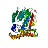
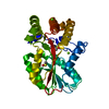
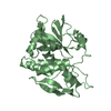
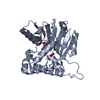
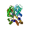
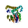
- PDB-7q2c: mycolic acid methyltransferase Hma (MmaA4) from Mycobac-terium tu... -
+ Open data

- Basic information
| Entry | Database: PDB / ID: 7q2c | |||||||||
|---|---|---|---|---|---|---|---|---|---|---|
| Title | mycolic acid methyltransferase Hma (MmaA4) from Mycobac-terium tuberculosis in complex with ZT260 | |||||||||
 Components Components | Hydroxymycolate synthase MmaA4 | |||||||||
 Keywords Keywords | TRANSFERASE / mycolic acid methyltransferase mycobacterium tuberculosis fragment based ligand discovery | |||||||||
| Function / homology |  Function and homology information Function and homology informationcyclopropane-fatty-acyl-phospholipid synthase activity / mycolic acid biosynthetic process / S-adenosylmethionine-dependent methyltransferase activity / lipid biosynthetic process / peptidoglycan-based cell wall / Transferases; Transferring one-carbon groups; Methyltransferases / methyltransferase activity / methylation / plasma membrane Similarity search - Function | |||||||||
| Biological species |   Mycobacterium tuberculosis (bacteria) Mycobacterium tuberculosis (bacteria) | |||||||||
| Method |  X-RAY DIFFRACTION / SYNCHROTRON / FOURIER SYNTHESIS / Resolution: 1.9 Å X-RAY DIFFRACTION / SYNCHROTRON / FOURIER SYNTHESIS / Resolution: 1.9 Å | |||||||||
 Authors Authors | Maveyraud, L. / Galy, R. / Mourey, L. | |||||||||
| Funding support |  France, 2items France, 2items
| |||||||||
 Citation Citation | Journal: Pharmaceuticals / Year: 2021 Title: Fragment-Based Ligand Discovery Applied to the Mycolic Acid Methyltransferase Hma (MmaA4) from Mycobacterium tuberculosis : A Crystallographic and Molecular Modelling Study. Authors: Galy, R. / Ballereau, S. / Genisson, Y. / Mourey, L. / Plaquevent, J.C. / Maveyraud, L. | |||||||||
| History |
|
- Structure visualization
| Structure viewer | Molecule: MolmilJmol/JSmol |
|---|
- Downloads & links
-Download
| PDBx/mmCIF format | 7q2c.cif.gz | 135.9 KB | Display | PDBx/mmCIF format |
|---|---|---|---|---|
| PDB format | pdb7q2c.ent.gz | 104.4 KB | Display | PDB format |
| PDBx/mmJSON format | 7q2c.json.gz | Tree view | PDBx/mmJSON format | |
| Others |  Other downloads Other downloads |
-Validation report
| Arichive directory | https://data.pdbj.org/pub/pdb/validation_reports/q2/7q2cftp://data.pdbj.org/pub/pdb/validation_reports/q2/7q2c | HTTPS FTP |
|---|
-Related structure data
| Related structure data |  7q2bC  7q2dC 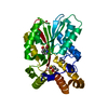 7q2eC  7q2fC  7q2gC 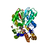 7q2hC  2fk7S C: citing same article ( S: Starting model for refinement |
|---|---|
| Similar structure data |


-Links
 PDBj
PDBj- Assembly
| Deposited unit | 
| ||||||||
|---|---|---|---|---|---|---|---|---|---|
| 1 |
| ||||||||
| Unit cell |
|
-Components
| #1: Protein | Mass: 36521.305 Da / Num. of mol.: 1 Source method: isolated from a genetically manipulated source Source: (gene. exp.) Mycobacterium tuberculosis (strain ATCC 25618 / H37Rv) (bacteria)Strain: ATCC 25618 / H37Rv / Gene: mmaA4, hma, mma4, Rv0642c / Plasmid: pET15b / Production host: References: UniProt: Q79FX8, Transferases; Transferring one-carbon groups; Methyltransferases | ||||||
|---|---|---|---|---|---|---|---|
| #2: Chemical |   Mass: 78.133 Da / Num. of mol.: 3 / Source method: obtained synthetically / Formula: C2H6OS / Comment: DMSO, precipitant*YM Mass: 78.133 Da / Num. of mol.: 3 / Source method: obtained synthetically / Formula: C2H6OS / Comment: DMSO, precipitant*YM#3: Chemical | 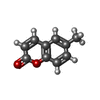  Mass: 160.169 Da / Num. of mol.: 2 / Source method: obtained synthetically / Formula: C10H8O2 / Feature type: SUBJECT OF INVESTIGATION Mass: 160.169 Da / Num. of mol.: 2 / Source method: obtained synthetically / Formula: C10H8O2 / Feature type: SUBJECT OF INVESTIGATION#4: Water | ChemComp-HOH / |  Mass: 18.015 Da / Num. of mol.: 138 / Source method: isolated from a natural source / Formula: H2O Mass: 18.015 Da / Num. of mol.: 138 / Source method: isolated from a natural source / Formula: H2OHas ligand of interest | Y | |
-Experimental details
-Experiment
| Experiment | Method: X-RAY DIFFRACTION / Number of used crystals: 1 |
|---|
- Sample preparation
| Crystal | Density Matthews: 2.65 Å3/Da / Density % sol: 53.66 % |
|---|---|
| Crystal grow | Temperature: 285 K / Method: vapor diffusion, sitting drop / pH: 6.5 / Details: BisTris 50 mM, PEG 3350 4% (w/v), pH 6.5 |
-Data collection
| Diffraction | Mean temperature: 100 K / Serial crystal experiment: N |
|---|---|
| Diffraction source | Source: SYNCHROTRON / Site: ESRF / Beamline: ID14-4 / Wavelength: 0.9393 Å |
| Detector | Type: ADSC QUANTUM 315r / Detector: CCD / Date: Oct 3, 2011 |
| Radiation | Protocol: SINGLE WAVELENGTH / Monochromatic (M) / Laue (L): M / Scattering type: x-ray |
| Radiation wavelength | Wavelength: 0.9393 Å / Relative weight: 1 |
| Reflection | Resolution: 1.85→35.66 Å / Num. obs: 33875 / % possible obs: 98.4 % / Redundancy: 5.11 % / CC1/2: 0.999 / Rrim(I) all: 0.056 / Rsym value: 0.051 / Net I/σ(I): 15.05 |
| Reflection shell | Resolution: 1.85→1.96 Å / Redundancy: 3.72 % / Mean I/σ(I) obs: 0.98 / Num. unique obs: 5254 / CC1/2: 0.591 / Rrim(I) all: 1.264 / Rsym value: 1.093 / % possible all: 96.7 |
- Processing
| Software |
| ||||||||||||||||||||||||||||||||||||||||||||||||||||||||||||||||||||||||||||||||||||||||||||||||||||||||||||||||||||||||||||||||||||||||||||||||||||||||||||||||||||||||||||||||||||||
|---|---|---|---|---|---|---|---|---|---|---|---|---|---|---|---|---|---|---|---|---|---|---|---|---|---|---|---|---|---|---|---|---|---|---|---|---|---|---|---|---|---|---|---|---|---|---|---|---|---|---|---|---|---|---|---|---|---|---|---|---|---|---|---|---|---|---|---|---|---|---|---|---|---|---|---|---|---|---|---|---|---|---|---|---|---|---|---|---|---|---|---|---|---|---|---|---|---|---|---|---|---|---|---|---|---|---|---|---|---|---|---|---|---|---|---|---|---|---|---|---|---|---|---|---|---|---|---|---|---|---|---|---|---|---|---|---|---|---|---|---|---|---|---|---|---|---|---|---|---|---|---|---|---|---|---|---|---|---|---|---|---|---|---|---|---|---|---|---|---|---|---|---|---|---|---|---|---|---|---|---|---|---|---|
| Refinement | Method to determine structure: FOURIER SYNTHESIS Starting model: 2FK7 Resolution: 1.9→35.69 Å / Cor.coef. Fo:Fc: 0.971 / Cor.coef. Fo:Fc free: 0.95 / SU B: 7.414 / SU ML: 0.102 / Cross valid method: THROUGHOUT / ESU R: 0.122 / ESU R Free: 0.126 / Stereochemistry target values: MAXIMUM LIKELIHOOD / Details: HYDROGENS HAVE BEEN ADDED IN THE RIDING POSITIONS
| ||||||||||||||||||||||||||||||||||||||||||||||||||||||||||||||||||||||||||||||||||||||||||||||||||||||||||||||||||||||||||||||||||||||||||||||||||||||||||||||||||||||||||||||||||||||
| Solvent computation | Ion probe radii: 0.8 Å / Shrinkage radii: 0.8 Å / VDW probe radii: 1.2 Å / Solvent model: MASK | ||||||||||||||||||||||||||||||||||||||||||||||||||||||||||||||||||||||||||||||||||||||||||||||||||||||||||||||||||||||||||||||||||||||||||||||||||||||||||||||||||||||||||||||||||||||
| Displacement parameters | Biso mean: 58.022 Å2
| ||||||||||||||||||||||||||||||||||||||||||||||||||||||||||||||||||||||||||||||||||||||||||||||||||||||||||||||||||||||||||||||||||||||||||||||||||||||||||||||||||||||||||||||||||||||
| Refinement step | Cycle: 1 / Resolution: 1.9→35.69 Å
| ||||||||||||||||||||||||||||||||||||||||||||||||||||||||||||||||||||||||||||||||||||||||||||||||||||||||||||||||||||||||||||||||||||||||||||||||||||||||||||||||||||||||||||||||||||||
| Refine LS restraints |
|