 Movie
Movie Controller

[English] 日本語
 Yorodumi
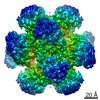
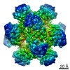
- PDB-7q07: Ketol-acid reductoisomerase from Methanothermococcus thermolithot... -
+ Open data

- Basic information
| Entry | Database: PDB / ID: 7q07 | ||||||
|---|---|---|---|---|---|---|---|
| Title | Ketol-acid reductoisomerase from Methanothermococcus thermolithotrophicus in the open state with NADP and tartrate | ||||||
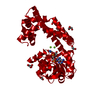
 Components Components | Ketol-Acid Reductoisomerase from Methanothermococcus thermolithotrophicus | ||||||
 Keywords Keywords | ISOMERASE / Ketol-acid reductoisomerase / KARI / methanogenic archaea / conformational rearrangement / native purification / oligomerization / thermophile / branched-chain amino acid / biosynthesis | ||||||
| Function / homology | NADP NICOTINAMIDE-ADENINE-DINUCLEOTIDE PHOSPHATE / L(+)-TARTARIC ACID Function and homology information Function and homology information | ||||||
| Biological species |  Methanothermococcus thermolithotrophicus DSM 2095 (archaea) Methanothermococcus thermolithotrophicus DSM 2095 (archaea) | ||||||
| Method |  X-RAY DIFFRACTION / SYNCHROTRON / MOLECULAR REPLACEMENT / Resolution: 2.2 Å X-RAY DIFFRACTION / SYNCHROTRON / MOLECULAR REPLACEMENT / Resolution: 2.2 Å | ||||||
 Authors Authors | Lemaire, O.N. / Mueller, M. / Wagner, T. | ||||||
| Funding support |  Germany, 1items Germany, 1items
| ||||||
 Citation Citation | Journal: Biomolecules / Year: 2021 Title: Structural Rearrangements of a Dodecameric Ketol-Acid Reductoisomerase Isolated from a Marine Thermophilic Methanogen. Authors: Lemaire, O.N. / Muller, M.C. / Kahnt, J. / Wagner, T. | ||||||
| History |
|
- Structure visualization
| Structure viewer | Molecule: MolmilJmol/JSmol |
|---|
- Downloads & links
-Download
| PDBx/mmCIF format | 7q07.cif.gz | 142.8 KB | Display | PDBx/mmCIF format |
|---|---|---|---|---|
| PDB format | pdb7q07.ent.gz | 110.9 KB | Display | PDB format |
| PDBx/mmJSON format | 7q07.json.gz | Tree view | PDBx/mmJSON format | |
| Others |  Other downloads Other downloads |
-Validation report
| Arichive directory | https://data.pdbj.org/pub/pdb/validation_reports/q0/7q07ftp://data.pdbj.org/pub/pdb/validation_reports/q0/7q07 | HTTPS FTP |
|---|
-Related structure data
| Related structure data |  7q03SC S: Starting model for refinement C: citing same article ( |
|---|---|
| Similar structure data |


-Links
 PDBj
PDBj
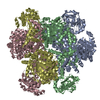
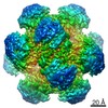
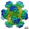
- Assembly
| Deposited unit | 
| |||||||||||||||
|---|---|---|---|---|---|---|---|---|---|---|---|---|---|---|---|---|
| 1 | x 12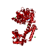
| |||||||||||||||
| Unit cell |
| |||||||||||||||
| Components on special symmetry positions |
|
-Components
-Protein , 1 types, 1 molecules A
| #1: Protein | Mass: 36556.746 Da / Num. of mol.: 1 / Source method: isolated from a natural source Source: (natural) Methanothermococcus thermolithotrophicus DSM 2095 (archaea)Cell line: / / Organ: / / Plasmid details: / / Variant: / / Tissue: / / References: ketol-acid reductoisomerase (NADP+) |
|---|
-Non-polymers , 5 types, 146 molecules 


| #2: Chemical | ChemComp-TLA /  Mass: 150.087 Da / Num. of mol.: 1 / Source method: obtained synthetically / Formula: C4H6O6 Mass: 150.087 Da / Num. of mol.: 1 / Source method: obtained synthetically / Formula: C4H6O6 | ||||||
|---|---|---|---|---|---|---|---|
| #3: Chemical |  Mass: 35.453 Da / Num. of mol.: 3 / Source method: obtained synthetically / Formula: Cl Mass: 35.453 Da / Num. of mol.: 3 / Source method: obtained synthetically / Formula: Cl#4: Chemical | ChemComp-EDO / |  Mass: 62.068 Da / Num. of mol.: 1 / Source method: obtained synthetically / Formula: C2H6O2 Mass: 62.068 Da / Num. of mol.: 1 / Source method: obtained synthetically / Formula: C2H6O2#5: Chemical | ChemComp-NAP / |  Mass: 743.405 Da / Num. of mol.: 1 / Source method: obtained synthetically / Formula: C21H28N7O17P3 / Feature type: SUBJECT OF INVESTIGATION Mass: 743.405 Da / Num. of mol.: 1 / Source method: obtained synthetically / Formula: C21H28N7O17P3 / Feature type: SUBJECT OF INVESTIGATION#6: Water | ChemComp-HOH / | Mass: 18.015 Da / Num. of mol.: 140 / Source method: isolated from a natural source / Formula: H2O |
-Details
| Has ligand of interest | Y |
|---|
-Experimental details
-Experiment
| Experiment | Method: X-RAY DIFFRACTION / Number of used crystals: 1 |
|---|
- Sample preparation
| Crystal | Density Matthews: 2.56 Å3/Da / Density % sol: 51.9 % / Description: Flower-shaped plates. |
|---|---|
| Crystal grow | Temperature: 291.15 K / Method: vapor diffusion, sitting drop / pH: 6.5 Details: KARI was crystallized at 13.5 mg/ml in the following buffer 25 mM Tris/HCl pH 7.6, 2 mM dithiothreitol, 10% glycerol. The reservoir was filled with 90 ul crystallization solution (33 % (w/v) ...Details: KARI was crystallized at 13.5 mg/ml in the following buffer 25 mM Tris/HCl pH 7.6, 2 mM dithiothreitol, 10% glycerol. The reservoir was filled with 90 ul crystallization solution (33 % (w/v) PEG 5000 MME, 100 mM MES/NaOH pH 6.5 and 200 mM ammonium sulphate). Drops of 0.7 ul protein with 0.7 ul of crystallisation solution were applied on the shelf. Crystals were firstly soaked in the crystallisation solution supplemented with 20 mM NADP, 50 mM L-Tartrate and 50 mM MgCl2 for 3 minutes, and then soaked in the crystallisation solution supplemented with 30 % v/v glycerol before freezing. Temp details: +/- 3 degree |
-Data collection
| Diffraction | Mean temperature: 100 K / Serial crystal experiment: N |
|---|---|
| Diffraction source | Source: SYNCHROTRON / Site: SLS  / Beamline: X10SA / Wavelength: 0.97916 Å / Beamline: X10SA / Wavelength: 0.97916 Å |
| Detector | Type: DECTRIS PILATUS 6M / Detector: PIXEL / Date: Jun 24, 2016 |
| Radiation | Protocol: SINGLE WAVELENGTH / Monochromatic (M) / Laue (L): M / Scattering type: x-ray |
| Radiation wavelength | Wavelength: 0.97916 Å / Relative weight: 1 |
| Reflection twin | Operator: -l,-k,-h / Fraction: 0.12 |
| Reflection | Resolution: 2.2→46.25 Å / Num. obs: 19073 / % possible obs: 100 % / Redundancy: 18.2 % / CC1/2: 0.998 / Rmerge(I) obs: 0.163 / Rpim(I) all: 0.039 / Rrim(I) all: 0.168 / Net I/σ(I): 23.7 |
| Reflection shell | Resolution: 2.2→2.32 Å / Redundancy: 10.9 % / Rmerge(I) obs: 0.528 / Mean I/σ(I) obs: 4.4 / Num. unique obs: 2766 / CC1/2: 0.385 / Rpim(I) all: 0.166 / Rrim(I) all: 0.554 / % possible all: 100 |
- Processing
| Software |
| |||||||||||||||||||||||||||||||||||||||||||||||||||||||||||||||||||||||||||
|---|---|---|---|---|---|---|---|---|---|---|---|---|---|---|---|---|---|---|---|---|---|---|---|---|---|---|---|---|---|---|---|---|---|---|---|---|---|---|---|---|---|---|---|---|---|---|---|---|---|---|---|---|---|---|---|---|---|---|---|---|---|---|---|---|---|---|---|---|---|---|---|---|---|---|---|---|
| Refinement | Method to determine structure: MOLECULAR REPLACEMENT Starting model: 7Q03 Resolution: 2.2→41.37 Å / Cross valid method: THROUGHOUT / σ(F): 133.59 / Phase error: 21.21 / Stereochemistry target values: TWIN_LSQ_F Details: The refinement was performed with TLS and intensity-based twin refinement with the following twin law -l,-k,-h
| |||||||||||||||||||||||||||||||||||||||||||||||||||||||||||||||||||||||||||
| Solvent computation | Shrinkage radii: 0.9 Å / VDW probe radii: 1.11 Å / Solvent model: FLAT BULK SOLVENT MODEL | |||||||||||||||||||||||||||||||||||||||||||||||||||||||||||||||||||||||||||
| Displacement parameters | Biso max: 98.42 Å2 / Biso mean: 30.3423 Å2 / Biso min: 9.07 Å2 | |||||||||||||||||||||||||||||||||||||||||||||||||||||||||||||||||||||||||||
| Refinement step | Cycle: final / Resolution: 2.2→41.37 Å
| |||||||||||||||||||||||||||||||||||||||||||||||||||||||||||||||||||||||||||
| LS refinement shell | Refine-ID: X-RAY DIFFRACTION / Rfactor Rfree error: 0 / Total num. of bins used: 7
| |||||||||||||||||||||||||||||||||||||||||||||||||||||||||||||||||||||||||||
| Refinement TLS params. | Method: refined / Refine-ID: X-RAY DIFFRACTION
| |||||||||||||||||||||||||||||||||||||||||||||||||||||||||||||||||||||||||||
| Refinement TLS group |
|
