 Movie
Movie Controller

[English] 日本語
 Yorodumi
- PDB-7q03: Ketol-acid reductoisomerase from Methanothermococcus thermolithot... -
+ Open data

- Basic information
| Entry | Database: PDB / ID: 7q03 | ||||||
|---|---|---|---|---|---|---|---|
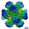
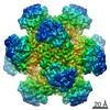
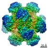
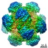
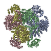
| Title | Ketol-acid reductoisomerase from Methanothermococcus thermolithotrophicus in the close state with NADP and Mg2+ | ||||||
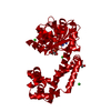
 Components Components | Ketol-acid reductoisomerase from Methanothermococcus thermolithotrophicus | ||||||
 Keywords Keywords | ISOMERASE / Ketol-acid reductoisomerase / KARI / methanogenic archaea / conformational rearrangement / native purification / oligomerization / thermophile / branched-chain keto acid / biosynthesis | ||||||
| Function / homology | NADP NICOTINAMIDE-ADENINE-DINUCLEOTIDE PHOSPHATE / 2-[2-(2-METHOXY-ETHOXY)-ETHOXY]-ETHOXYL Function and homology information Function and homology information | ||||||
| Biological species |  Methanothermococcus thermolithotrophicus DSM 2095 (archaea) Methanothermococcus thermolithotrophicus DSM 2095 (archaea) | ||||||
| Method |  X-RAY DIFFRACTION / SYNCHROTRON / MOLECULAR REPLACEMENT / Resolution: 2.1 Å X-RAY DIFFRACTION / SYNCHROTRON / MOLECULAR REPLACEMENT / Resolution: 2.1 Å | ||||||
 Authors Authors | Lemaire, O.N. / Mueller, M. / Wagner, T. | ||||||
| Funding support |  Germany, 1items Germany, 1items
| ||||||
 Citation Citation | Journal: Biomolecules / Year: 2021 Title: Structural Rearrangements of a Dodecameric Ketol-Acid Reductoisomerase Isolated from a Marine Thermophilic Methanogen. Authors: Lemaire, O.N. / Muller, M.C. / Kahnt, J. / Wagner, T. | ||||||
| History |
|
- Structure visualization
| Structure viewer | Molecule: MolmilJmol/JSmol |
|---|
- Downloads & links
-Download
| PDBx/mmCIF format | 7q03.cif.gz | 145.5 KB | Display | PDBx/mmCIF format |
|---|---|---|---|---|
| PDB format | pdb7q03.ent.gz | 113.3 KB | Display | PDB format |
| PDBx/mmJSON format | 7q03.json.gz | Tree view | PDBx/mmJSON format | |
| Others |  Other downloads Other downloads |
-Validation report
| Arichive directory | https://data.pdbj.org/pub/pdb/validation_reports/q0/7q03ftp://data.pdbj.org/pub/pdb/validation_reports/q0/7q03 | HTTPS FTP |
|---|
-Related structure data
| Related structure data |  7q07C 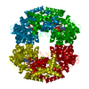 4tskS S: Starting model for refinement C: citing same article ( |
|---|---|
| Similar structure data |


-Links
 PDBj
PDBj- Assembly
| Deposited unit | 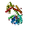
| ||||||||
|---|---|---|---|---|---|---|---|---|---|
| 1 | x 12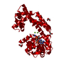
| ||||||||
| Unit cell |
|
-Components
-Protein , 1 types, 1 molecules A
| #1: Protein | Mass: 36556.746 Da / Num. of mol.: 1 / Source method: isolated from a natural source Source: (natural) Methanothermococcus thermolithotrophicus DSM 2095 (archaea)Cell line: / / Organ: / / Plasmid details: / / Variant: / / Tissue: / / References: ketol-acid reductoisomerase (NADP+) |
|---|
-Non-polymers , 7 types, 74 molecules 


| #2: Chemical |  Mass: 92.094 Da / Num. of mol.: 3 / Source method: obtained synthetically / Formula: C3H8O3 Mass: 92.094 Da / Num. of mol.: 3 / Source method: obtained synthetically / Formula: C3H8O3#3: Chemical |  Mass: 62.068 Da / Num. of mol.: 3 / Source method: obtained synthetically / Formula: C2H6O2 Mass: 62.068 Da / Num. of mol.: 3 / Source method: obtained synthetically / Formula: C2H6O2#4: Chemical | ChemComp-TOE / |  Mass: 164.200 Da / Num. of mol.: 1 / Source method: obtained synthetically / Formula: C7H16O4 Mass: 164.200 Da / Num. of mol.: 1 / Source method: obtained synthetically / Formula: C7H16O4#5: Chemical | ChemComp-NAP / |  Mass: 743.405 Da / Num. of mol.: 1 / Source method: obtained synthetically / Formula: C21H28N7O17P3 / Feature type: SUBJECT OF INVESTIGATION Mass: 743.405 Da / Num. of mol.: 1 / Source method: obtained synthetically / Formula: C21H28N7O17P3 / Feature type: SUBJECT OF INVESTIGATION#6: Chemical |  Mass: 24.305 Da / Num. of mol.: 2 / Source method: obtained synthetically / Formula: Mg / Feature type: SUBJECT OF INVESTIGATION Mass: 24.305 Da / Num. of mol.: 2 / Source method: obtained synthetically / Formula: Mg / Feature type: SUBJECT OF INVESTIGATION#7: Chemical | ChemComp-SO4 / |  Mass: 96.063 Da / Num. of mol.: 1 / Source method: obtained synthetically / Formula: SO4 Mass: 96.063 Da / Num. of mol.: 1 / Source method: obtained synthetically / Formula: SO4#8: Water | ChemComp-HOH / | Mass: 18.015 Da / Num. of mol.: 63 / Source method: isolated from a natural source / Formula: H2O |
|---|
-Details
| Has ligand of interest | Y |
|---|
-Experimental details
-Experiment
| Experiment | Method: X-RAY DIFFRACTION / Number of used crystals: 1 |
|---|
- Sample preparation
| Crystal | Density Matthews: 2.5 Å3/Da / Density % sol: 50.84 % / Description: Transparent plates |
|---|---|
| Crystal grow | Temperature: 291.15 K / Method: vapor diffusion, sitting drop / pH: 8 Details: The reservoir was filled with 90 ul of crystallisation solution: 20% (w/v) PEG 6000, 100 mM Tris/HCl pH 8.0 and 200 mM lithium chloride. The protein was crystallized at 13 mg/ml in the ...Details: The reservoir was filled with 90 ul of crystallisation solution: 20% (w/v) PEG 6000, 100 mM Tris/HCl pH 8.0 and 200 mM lithium chloride. The protein was crystallized at 13 mg/ml in the following buffer 25 mM Tris/HCl pH 7.6, 2 mM dithiothreitol, 10% (v/v) glycerol. Drops of 0.7 ul protein with 0.7 ul of crystallisation solution were applied on the shelf. Crystals were soaked in the crystallisation solution supplemented with 30% (v/v) ethylene glycol for few seconds before freezing in liquid nitrogen. Temp details: +/- 3 degrees |
-Data collection
| Diffraction | Mean temperature: 100 K / Serial crystal experiment: N |
|---|---|
| Diffraction source | Source: SYNCHROTRON / Site: SLS  / Beamline: X10SA / Wavelength: 1.73913 Å / Beamline: X10SA / Wavelength: 1.73913 Å |
| Detector | Type: DECTRIS PILATUS 6M / Detector: PIXEL / Date: Nov 30, 2015 |
| Radiation | Protocol: SINGLE WAVELENGTH / Monochromatic (M) / Laue (L): M / Scattering type: x-ray |
| Radiation wavelength | Wavelength: 1.73913 Å / Relative weight: 1 |
| Reflection twin | Operator: -l,-k,-h / Fraction: 0.32 |
| Reflection | Resolution: 2.1→45.92 Å / Num. obs: 21408 / % possible obs: 100 % / Redundancy: 14.3 % / CC1/2: 0.999 / Rmerge(I) obs: 0.095 / Rpim(I) all: 0.026 / Rrim(I) all: 0.098 / Net I/σ(I): 18.3 |
| Reflection shell | Resolution: 2.1→2.21 Å / Redundancy: 13.3 % / Rmerge(I) obs: 0.744 / Mean I/σ(I) obs: 3.5 / Num. unique obs: 3090 / CC1/2: 0.468 / Rpim(I) all: 0.212 / Rrim(I) all: 0.774 / % possible all: 100 |
- Processing
| Software |
| ||||||||||||||||||||||||||||||||||||||||||||||||||||||||||||||||||||||||||||||||||||||||||||||||||||
|---|---|---|---|---|---|---|---|---|---|---|---|---|---|---|---|---|---|---|---|---|---|---|---|---|---|---|---|---|---|---|---|---|---|---|---|---|---|---|---|---|---|---|---|---|---|---|---|---|---|---|---|---|---|---|---|---|---|---|---|---|---|---|---|---|---|---|---|---|---|---|---|---|---|---|---|---|---|---|---|---|---|---|---|---|---|---|---|---|---|---|---|---|---|---|---|---|---|---|---|---|---|
| Refinement | Method to determine structure: MOLECULAR REPLACEMENT Starting model: 4TSK Resolution: 2.1→41.07 Å / Cross valid method: THROUGHOUT / σ(F): 39.04 / Phase error: 30.48 / Stereochemistry target values: TWIN_LSQ_F Details: The refinement was performed with TLS and using intensity-based twin refinement with the following twin law -l,-k,-h
| ||||||||||||||||||||||||||||||||||||||||||||||||||||||||||||||||||||||||||||||||||||||||||||||||||||
| Solvent computation | Shrinkage radii: 0.9 Å / VDW probe radii: 1.11 Å / Solvent model: FLAT BULK SOLVENT MODEL | ||||||||||||||||||||||||||||||||||||||||||||||||||||||||||||||||||||||||||||||||||||||||||||||||||||
| Displacement parameters | Biso max: 129.52 Å2 / Biso mean: 45.8799 Å2 / Biso min: 23.36 Å2 | ||||||||||||||||||||||||||||||||||||||||||||||||||||||||||||||||||||||||||||||||||||||||||||||||||||
| Refinement step | Cycle: final / Resolution: 2.1→41.07 Å
| ||||||||||||||||||||||||||||||||||||||||||||||||||||||||||||||||||||||||||||||||||||||||||||||||||||
| LS refinement shell | Refine-ID: X-RAY DIFFRACTION / Rfactor Rfree error: 0 / Total num. of bins used: 8
| ||||||||||||||||||||||||||||||||||||||||||||||||||||||||||||||||||||||||||||||||||||||||||||||||||||
| Refinement TLS params. | Method: refined / Refine-ID: X-RAY DIFFRACTION
| ||||||||||||||||||||||||||||||||||||||||||||||||||||||||||||||||||||||||||||||||||||||||||||||||||||
| Refinement TLS group |
|
