 Movie
Movie Controller

[English] 日本語
 Yorodumi
- PDB-7q02: Zn-free structure of lipocalin-like Milk protein, inspired from D... -
+ Open data

- Basic information
| Entry | Database: PDB / ID: 7q02 | ||||||||||||
|---|---|---|---|---|---|---|---|---|---|---|---|---|---|
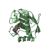
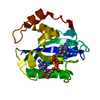
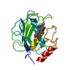
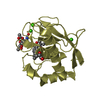
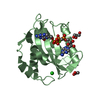
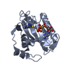
| Title | Zn-free structure of lipocalin-like Milk protein, inspired from Diploptera punctata, expressed in Saccharomyces cerevisiae | ||||||||||||
 Components Components | Milk protein | ||||||||||||
 Keywords Keywords | LIPID BINDING PROTEIN / Lipocalin / Milk Protein / Dipolptera punctata | ||||||||||||
| Function / homology | Calycin / metal ion binding / PALMITOLEIC ACID / Milk protein Function and homology information Function and homology information | ||||||||||||
| Biological species |  Diploptera punctata (Pacific beetle cockroach) Diploptera punctata (Pacific beetle cockroach) | ||||||||||||
| Method |  X-RAY DIFFRACTION / SYNCHROTRON / MOLECULAR REPLACEMENT / Resolution: 1.45 Å X-RAY DIFFRACTION / SYNCHROTRON / MOLECULAR REPLACEMENT / Resolution: 1.45 Å | ||||||||||||
 Authors Authors | Banerjee, S. / Dhanabalan, K.V. / Ramaswamy, S. | ||||||||||||
| Funding support |  India, 3items India, 3items
| ||||||||||||
 Citation Citation | Journal: Biochim Biophys Acta Gen Subj / Year: 2022 Title: Structure of recombinantly expressed cockroach Lili-Mip protein in glycosylated and deglycosylated forms. Authors: KanagaVijayan, D. / Subramanian, R. / Santhakumari, P.R. / Chavas, L.M.G. / Banerjee, S. | ||||||||||||
| History |
|
- Structure visualization
| Structure viewer | Molecule: MolmilJmol/JSmol |
|---|
- Downloads & links
-Download
| PDBx/mmCIF format | 7q02.cif.gz | 88.3 KB | Display | PDBx/mmCIF format |
|---|---|---|---|---|
| PDB format | pdb7q02.ent.gz | 63.8 KB | Display | PDB format |
| PDBx/mmJSON format | 7q02.json.gz | Tree view | PDBx/mmJSON format | |
| Others |  Other downloads Other downloads |
-Validation report
| Arichive directory | https://data.pdbj.org/pub/pdb/validation_reports/q0/7q02ftp://data.pdbj.org/pub/pdb/validation_reports/q0/7q02 | HTTPS FTP |
|---|
-Related structure data
| Related structure data |  7bkxC  4nyqS C: citing same article ( S: Starting model for refinement |
|---|---|
| Similar structure data |


-Links
 PDBj
PDBj

- Assembly
| Deposited unit | 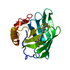
| ||||||||
|---|---|---|---|---|---|---|---|---|---|
| 1 |
| ||||||||
| Unit cell |
|
-Components
| #1: Protein | Mass: 18853.268 Da / Num. of mol.: 1 Source method: isolated from a genetically manipulated source Source: (gene. exp.) Diploptera punctata (Pacific beetle cockroach)Production host:  |
|---|---|
| #2: Chemical | ChemComp-PAM /   Mass: 254.408 Da / Num. of mol.: 1 / Source method: obtained synthetically / Formula: C16H30O2 / Feature type: SUBJECT OF INVESTIGATION Mass: 254.408 Da / Num. of mol.: 1 / Source method: obtained synthetically / Formula: C16H30O2 / Feature type: SUBJECT OF INVESTIGATION |
| #3: Water | ChemComp-HOH /  Mass: 18.015 Da / Num. of mol.: 224 / Source method: isolated from a natural source / Formula: H2O Mass: 18.015 Da / Num. of mol.: 224 / Source method: isolated from a natural source / Formula: H2O |
| Has ligand of interest | Y |
| Has protein modification | Y |
-Experimental details
-Experiment
| Experiment | Method: X-RAY DIFFRACTION / Number of used crystals: 1 |
|---|
- Sample preparation
| Crystal | Density Matthews: 2.17 Å3/Da / Density % sol: 43.26 % / Description: Plate-like crystals. |
|---|---|
| Crystal grow | Temperature: 290 K / Method: vapor diffusion, hanging drop / pH: 4.5 Details: 0.1 M Sodium Acetate, HCl, pH 4.5; 25 % (w/v) PEG 3350 Temp details: Controlled room |
-Data collection
| Diffraction | Mean temperature: 100 K / Ambient temp details: 100 / Serial crystal experiment: N | ||||||||||||||||||||||||||||||
|---|---|---|---|---|---|---|---|---|---|---|---|---|---|---|---|---|---|---|---|---|---|---|---|---|---|---|---|---|---|---|---|
| Diffraction source | Source: SYNCHROTRON / Site: APS  / Beamline: 23-ID-D / Wavelength: 1.0332 Å / Beamline: 23-ID-D / Wavelength: 1.0332 Å | ||||||||||||||||||||||||||||||
| Detector | Type: DECTRIS PILATUS 6M / Detector: PIXEL / Date: Oct 9, 2021 | ||||||||||||||||||||||||||||||
| Radiation | Protocol: SINGLE WAVELENGTH / Monochromatic (M) / Laue (L): M / Scattering type: x-ray | ||||||||||||||||||||||||||||||
| Radiation wavelength | Wavelength: 1.0332 Å / Relative weight: 1 | ||||||||||||||||||||||||||||||
| Reflection | Resolution: 1.45→48.81 Å / Num. obs: 28438 / % possible obs: 100 % / Redundancy: 6.3 % / Biso Wilson estimate: 12.79 Å2 / CC1/2: 0.997 / Rmerge(I) obs: 0.107 / Rpim(I) all: 0.046 / Rrim(I) all: 0.117 / Net I/σ(I): 10.7 / Num. measured all: 180115 / Scaling rejects: 137 | ||||||||||||||||||||||||||||||
| Reflection shell | Diffraction-ID: 1
|
- Processing
| Software |
| ||||||||||||||||||||||||||||||||||||||||||||||||||||||||||||||||||
|---|---|---|---|---|---|---|---|---|---|---|---|---|---|---|---|---|---|---|---|---|---|---|---|---|---|---|---|---|---|---|---|---|---|---|---|---|---|---|---|---|---|---|---|---|---|---|---|---|---|---|---|---|---|---|---|---|---|---|---|---|---|---|---|---|---|---|---|
| Refinement | Method to determine structure: MOLECULAR REPLACEMENT Starting model: 4NYQ Resolution: 1.45→48.81 Å / SU ML: 0.13 / Cross valid method: THROUGHOUT / σ(F): 1.34 / Phase error: 18.32 / Stereochemistry target values: ML
| ||||||||||||||||||||||||||||||||||||||||||||||||||||||||||||||||||
| Solvent computation | Shrinkage radii: 0.9 Å / VDW probe radii: 1.11 Å / Solvent model: FLAT BULK SOLVENT MODEL | ||||||||||||||||||||||||||||||||||||||||||||||||||||||||||||||||||
| Displacement parameters | Biso max: 59.2 Å2 / Biso mean: 18.435 Å2 / Biso min: 7.62 Å2 | ||||||||||||||||||||||||||||||||||||||||||||||||||||||||||||||||||
| Refinement step | Cycle: final / Resolution: 1.45→48.81 Å
| ||||||||||||||||||||||||||||||||||||||||||||||||||||||||||||||||||
| LS refinement shell | Refine-ID: X-RAY DIFFRACTION / Rfactor Rfree error: 0 / Total num. of bins used: 10 / % reflection obs: 100 %
|
