 Movie
Movie Controller


+ Open data

- Basic information
| Entry | Database: PDB / ID: 7pyi | |||||||||
|---|---|---|---|---|---|---|---|---|---|---|
| Title | Structure of LPMO in complex with cellotetraose at 6.65x10^6 Gy | |||||||||
 Components Components | Auxiliary activity 9 | |||||||||
 Keywords Keywords | OXIDOREDUCTASE / lytic polysaccharide monooxygenase / metalloenzyme / AA9 | |||||||||
| Function / homology |  Function and homology information Function and homology informationlytic cellulose monooxygenase (C4-dehydrogenating) / cellulose catabolic process / monooxygenase activity / extracellular region / metal ion binding Similarity search - Function | |||||||||
| Biological species |  Lentinus similis (fungus) Lentinus similis (fungus) | |||||||||
| Method |  X-RAY DIFFRACTION / SYNCHROTRON / MOLECULAR REPLACEMENT / Resolution: 2.05 Å X-RAY DIFFRACTION / SYNCHROTRON / MOLECULAR REPLACEMENT / Resolution: 2.05 Å | |||||||||
 Authors Authors | Tandrup, T. / Lo Leggio, L. | |||||||||
| Funding support |  Denmark, 2items Denmark, 2items
| |||||||||
 Citation Citation | Journal: Iucrj / Year: 2022 Title: Changes in active-site geometry on X-ray photoreduction of a lytic polysaccharide monooxygenase active-site copper and saccharide binding. Authors: Tandrup, T. / Muderspach, S.J. / Banerjee, S. / Santoni, G. / Ipsen, J.O. / Hernandez-Rollan, C. / Norholm, M.H.H. / Johansen, K.S. / Meilleur, F. / Lo Leggio, L. | |||||||||
| History |
|
- Structure visualization
| Structure viewer | Molecule: MolmilJmol/JSmol |
|---|
- Downloads & links
-Download
| PDBx/mmCIF format | 7pyi.cif.gz | 65 KB | Display | PDBx/mmCIF format |
|---|---|---|---|---|
| PDB format | pdb7pyi.ent.gz | 46 KB | Display | PDB format |
| PDBx/mmJSON format | 7pyi.json.gz | Tree view | PDBx/mmJSON format | |
| Others |  Other downloads Other downloads |
-Validation report
| Arichive directory | https://data.pdbj.org/pub/pdb/validation_reports/py/7pyiftp://data.pdbj.org/pub/pdb/validation_reports/py/7pyi | HTTPS FTP |
|---|
-Related structure data
| Related structure data |  7pqrC 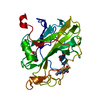 7pxiC 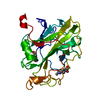 7pxjC  7pxkC 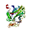 7pxlC 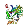 7pxmC 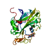 7pxnC 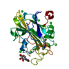 7pxrC  7pxsC  7pxtC 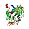 7pxuC  7pxvC 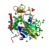 7pxwC 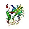 7pydC  7pyeC  7pyfC  7pygC 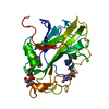 7pyhC 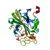 7pylC  7pymC 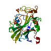 7pynC 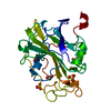 7pyoC  7pypC 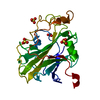 7pyqC 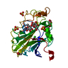 7pyuC  7pywC  7pyxC  7pyyC 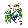 7pyzC  7pz0C 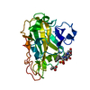 7pz3C 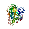 7pz4C 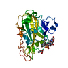 7pz5C 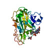 7pz6C  7pz7C  7pz8C  5achS C: citing same article ( S: Starting model for refinement |
|---|---|
| Similar structure data |
-Links
 PDBj
PDBj- Assembly
| Deposited unit | 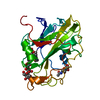
| |||||||||
|---|---|---|---|---|---|---|---|---|---|---|
| 1 |
| |||||||||
| Unit cell |
| |||||||||
| Components on special symmetry positions |
|
-Components
-Protein , 1 types, 1 molecules A
| #1: Protein | Mass: 25272.850 Da / Num. of mol.: 1 Source method: isolated from a genetically manipulated source Source: (gene. exp.) Lentinus similis (fungus) / Production host: |
|---|
-Sugars , 2 types, 2 molecules 
| #2: Polysaccharide | beta-D-glucopyranose-(1-4)-beta-D-glucopyranose-(1-4)-beta-D-glucopyranose-(1-4)-beta-D-glucopyranose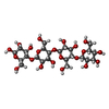  Source method: isolated from a genetically manipulated source Details: oligosaccharide / References: beta-cellotetraose |
|---|---|
| #4: Sugar | ChemComp-NAG /  Type: D-saccharide, beta linking / Mass: 221.208 Da / Num. of mol.: 1 / Source method: obtained synthetically / Formula: C8H15NO6 Type: D-saccharide, beta linking / Mass: 221.208 Da / Num. of mol.: 1 / Source method: obtained synthetically / Formula: C8H15NO6 |
-Non-polymers , 3 types, 67 molecules 


| #3: Chemical | ChemComp-CU /  Mass: 63.546 Da / Num. of mol.: 1 / Source method: obtained synthetically / Formula: Cu / Feature type: SUBJECT OF INVESTIGATION Mass: 63.546 Da / Num. of mol.: 1 / Source method: obtained synthetically / Formula: Cu / Feature type: SUBJECT OF INVESTIGATION |
|---|---|
| #5: Chemical | ChemComp-CL /  Mass: 35.453 Da / Num. of mol.: 1 / Source method: obtained synthetically / Formula: Cl / Feature type: SUBJECT OF INVESTIGATION Mass: 35.453 Da / Num. of mol.: 1 / Source method: obtained synthetically / Formula: Cl / Feature type: SUBJECT OF INVESTIGATION |
| #6: Water | ChemComp-HOH / Mass: 18.015 Da / Num. of mol.: 65 / Source method: isolated from a natural source / Formula: H2O |
-Details
| Has ligand of interest | Y |
|---|
-Experimental details
-Experiment
| Experiment | Method: X-RAY DIFFRACTION / Number of used crystals: 1 |
|---|
- Sample preparation
| Crystal | Density Matthews: 3.3 Å3/Da / Density % sol: 62.71 % |
|---|---|
| Crystal grow | Temperature: 298 K / Method: vapor diffusion, sitting drop / pH: 3.5 / Details: 3.3 M NaCl, 0.1 M Citric acid pH 3.5 |
-Data collection
| Diffraction | Mean temperature: 100 K / Serial crystal experiment: N |
|---|---|
| Diffraction source | Source: SYNCHROTRON / Site: PETRA III, DESY  / Beamline: P11 / Wavelength: 1.033 Å / Beamline: P11 / Wavelength: 1.033 Å |
| Detector | Type: DECTRIS EIGER2 X 16M / Detector: PIXEL / Date: Sep 29, 2016 |
| Radiation | Protocol: SINGLE WAVELENGTH / Monochromatic (M) / Laue (L): M / Scattering type: x-ray |
| Radiation wavelength | Wavelength: 1.033 Å / Relative weight: 1 |
| Reflection | Resolution: 2.05→89.1 Å / Num. obs: 21166 / % possible obs: 95.9 % / Redundancy: 3.29 % / CC1/2: 0.99 / Net I/σ(I): 15.87 |
| Reflection shell | Resolution: 2.05→2.1 Å / Num. unique obs: 1596 / CC1/2: 0.48 |
- Processing
| Software |
| ||||||||||||||||||||||||
|---|---|---|---|---|---|---|---|---|---|---|---|---|---|---|---|---|---|---|---|---|---|---|---|---|---|
| Refinement | Method to determine structure: MOLECULAR REPLACEMENT Starting model: 5ACH Resolution: 2.05→89.1 Å / Cor.coef. Fo:Fc: 0.964 / Cor.coef. Fo:Fc free: 0.949 / Cross valid method: THROUGHOUT / σ(F): 0 / Stereochemistry target values: MAXIMUM LIKELIHOOD
| ||||||||||||||||||||||||
| Solvent computation | Solvent model: MASK | ||||||||||||||||||||||||
| Displacement parameters | Biso max: 165.51 Å2 / Biso mean: 53.726 Å2 / Biso min: 28.38 Å2 | ||||||||||||||||||||||||
| Refinement step | Cycle: final / Resolution: 2.05→89.1 Å
| ||||||||||||||||||||||||
| LS refinement shell | Resolution: 2.05→2.103 Å
|
