 Movie
Movie Controller


+ Open data

- Basic information
| Entry | Database: PDB / ID: 7pwj | ||||||
|---|---|---|---|---|---|---|---|
| Title | dUTPase from human in complex with Stl | ||||||
 Components Components |
| ||||||
 Keywords Keywords | STRUCTURAL PROTEIN / dUTPase / dUTP / human / DNA | ||||||
| Function / homology |  Function and homology information Function and homology informationpyrimidine deoxyribonucleotide binding / dUTP catabolic process / dUMP biosynthetic process / dUTP diphosphatase / dUTP diphosphatase activity / signaling receptor inhibitor activity / Interconversion of nucleotide di- and triphosphates / peroxisome proliferator activated receptor binding / nucleobase-containing compound metabolic process / dTMP biosynthetic process ...pyrimidine deoxyribonucleotide binding / dUTP catabolic process / dUMP biosynthetic process / dUTP diphosphatase / dUTP diphosphatase activity / signaling receptor inhibitor activity / Interconversion of nucleotide di- and triphosphates / peroxisome proliferator activated receptor binding / nucleobase-containing compound metabolic process / dTMP biosynthetic process / regulation of protein-containing complex assembly / liver development / DNA replication / magnesium ion binding / mitochondrion / DNA binding / RNA binding / extracellular exosome / nucleoplasm / identical protein binding / nucleus Similarity search - Function | ||||||
| Biological species |   Staphylococcus aureus (bacteria) Staphylococcus aureus (bacteria) Homo sapiens (human) Homo sapiens (human) | ||||||
| Method |  X-RAY DIFFRACTION / SYNCHROTRON / MOLECULAR REPLACEMENT / Resolution: 1.944 Å X-RAY DIFFRACTION / SYNCHROTRON / MOLECULAR REPLACEMENT / Resolution: 1.944 Å | ||||||
 Authors Authors | Sanz-Frasquet, C. / Marina, A. | ||||||
| Funding support |  Spain, 1items Spain, 1items
| ||||||
 Citation Citation | Journal: Microbiol Spectr / Year: 2023 Title: The Bacteriophage-Phage-Inducible Chromosomal Island Arms Race Designs an Interkingdom Inhibitor of dUTPases. Authors: Sanz-Frasquet, C. / Ciges-Tomas, J.R. / Alite, C. / Penades, J.R. / Marina, A. | ||||||
| History |
|
- Structure visualization
| Structure viewer | Molecule: MolmilJmol/JSmol |
|---|
- Downloads & links
-Download
| PDBx/mmCIF format | 7pwj.cif.gz | 193 KB | Display | PDBx/mmCIF format |
|---|---|---|---|---|
| PDB format | pdb7pwj.ent.gz | Display | PDB format | |
| PDBx/mmJSON format | 7pwj.json.gz | Tree view | PDBx/mmJSON format | |
| Others |  Other downloads Other downloads |
-Validation report
| Arichive directory | https://data.pdbj.org/pub/pdb/validation_reports/pw/7pwjftp://data.pdbj.org/pub/pdb/validation_reports/pw/7pwj | HTTPS FTP |
|---|
-Related structure data
| Related structure data |  7pwxC 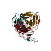 1q5uS 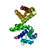 6h49S S: Starting model for refinement C: citing same article ( |
|---|---|
| Similar structure data |
-Links
 PDBj
PDBj


- Assembly
| Deposited unit | 
| ||||||||
|---|---|---|---|---|---|---|---|---|---|
| 1 |
| ||||||||
| Unit cell |
|
-Components
-Protein , 2 types, 6 molecules CCCEEEFFFAAABBBDDD
| #1: Protein | Mass: 18100.340 Da / Num. of mol.: 3 Source method: isolated from a genetically manipulated source Source: (gene. exp.) Staphylococcus aureus (bacteria) / Plasmid: pETNKI-1.10 / Production host: #2: Protein | Mass: 15910.923 Da / Num. of mol.: 3 Source method: isolated from a genetically manipulated source Source: (gene. exp.) Homo sapiens (human) / Gene: DUT / Production host: |
|---|
-Non-polymers , 4 types, 347 molecules 


| #3: Chemical | ChemComp-EDO /  Mass: 62.068 Da / Num. of mol.: 20 / Source method: obtained synthetically / Formula: C2H6O2 Mass: 62.068 Da / Num. of mol.: 20 / Source method: obtained synthetically / Formula: C2H6O2#4: Chemical | ChemComp-SO4 /  Mass: 96.063 Da / Num. of mol.: 6 / Source method: obtained synthetically / Formula: SO4 Mass: 96.063 Da / Num. of mol.: 6 / Source method: obtained synthetically / Formula: SO4#5: Chemical | ChemComp-GOL /  Mass: 92.094 Da / Num. of mol.: 6 / Source method: obtained synthetically / Formula: C3H8O3 Mass: 92.094 Da / Num. of mol.: 6 / Source method: obtained synthetically / Formula: C3H8O3#6: Water | ChemComp-HOH / | Mass: 18.015 Da / Num. of mol.: 315 / Source method: isolated from a natural source / Formula: H2O |
|---|
-Details
| Has ligand of interest | N |
|---|
-Experimental details
-Experiment
| Experiment | Method: X-RAY DIFFRACTION / Number of used crystals: 1 |
|---|
- Sample preparation
| Crystal | Density Matthews: 3.33 Å3/Da / Density % sol: 63.02 % |
|---|---|
| Crystal grow | Temperature: 294 K / Method: vapor diffusion, sitting drop / Details: 1.6M ammonium sulphate and 1M lithium sulphate. |
-Data collection
| Diffraction | Mean temperature: 80 K / Serial crystal experiment: N |
|---|---|
| Diffraction source | Source: SYNCHROTRON / Site: Diamond  / Beamline: I24 / Wavelength: 0.99186 Å / Beamline: I24 / Wavelength: 0.99186 Å |
| Detector | Type: DECTRIS EIGER2 X 9M / Detector: PIXEL / Date: Nov 24, 2019 |
| Radiation | Protocol: SINGLE WAVELENGTH / Monochromatic (M) / Laue (L): M / Scattering type: x-ray |
| Radiation wavelength | Wavelength: 0.99186 Å / Relative weight: 1 |
| Reflection | Resolution: 1.944→99.2 Å / Num. obs: 92354 / % possible obs: 98.48 % / Redundancy: 12.9 % / CC1/2: 0.999 / Net I/σ(I): 18.9 |
| Reflection shell | Resolution: 1.944→1.995 Å / Num. unique obs: 5386 / CC1/2: 0.897 |
- Processing
| Software |
| ||||||||||||||||||||||||||||||||||||||||||||||||||||||||||||||||||||||||||||||||||||||||||||||||||||||||||||||||||||||||||||||||||||||||||||||||||||||||||||||||
|---|---|---|---|---|---|---|---|---|---|---|---|---|---|---|---|---|---|---|---|---|---|---|---|---|---|---|---|---|---|---|---|---|---|---|---|---|---|---|---|---|---|---|---|---|---|---|---|---|---|---|---|---|---|---|---|---|---|---|---|---|---|---|---|---|---|---|---|---|---|---|---|---|---|---|---|---|---|---|---|---|---|---|---|---|---|---|---|---|---|---|---|---|---|---|---|---|---|---|---|---|---|---|---|---|---|---|---|---|---|---|---|---|---|---|---|---|---|---|---|---|---|---|---|---|---|---|---|---|---|---|---|---|---|---|---|---|---|---|---|---|---|---|---|---|---|---|---|---|---|---|---|---|---|---|---|---|---|---|---|---|---|
| Refinement | Method to determine structure: MOLECULAR REPLACEMENT Starting model: 6H49, 1Q5U Resolution: 1.944→99.199 Å / Cor.coef. Fo:Fc: 0.963 / Cor.coef. Fo:Fc free: 0.945 / SU B: 7.503 / SU ML: 0.185 / Cross valid method: FREE R-VALUE / ESU R: 0.147 / ESU R Free: 0.148 Details: Hydrogens have been added in their riding positions
| ||||||||||||||||||||||||||||||||||||||||||||||||||||||||||||||||||||||||||||||||||||||||||||||||||||||||||||||||||||||||||||||||||||||||||||||||||||||||||||||||
| Solvent computation | Ion probe radii: 0.8 Å / Shrinkage radii: 0.8 Å / VDW probe radii: 1.2 Å / Solvent model: MASK BULK SOLVENT | ||||||||||||||||||||||||||||||||||||||||||||||||||||||||||||||||||||||||||||||||||||||||||||||||||||||||||||||||||||||||||||||||||||||||||||||||||||||||||||||||
| Displacement parameters | Biso mean: 48.965 Å2
| ||||||||||||||||||||||||||||||||||||||||||||||||||||||||||||||||||||||||||||||||||||||||||||||||||||||||||||||||||||||||||||||||||||||||||||||||||||||||||||||||
| Refinement step | Cycle: LAST / Resolution: 1.944→99.199 Å
| ||||||||||||||||||||||||||||||||||||||||||||||||||||||||||||||||||||||||||||||||||||||||||||||||||||||||||||||||||||||||||||||||||||||||||||||||||||||||||||||||
| Refine LS restraints |
| ||||||||||||||||||||||||||||||||||||||||||||||||||||||||||||||||||||||||||||||||||||||||||||||||||||||||||||||||||||||||||||||||||||||||||||||||||||||||||||||||
| LS refinement shell |
|
