 Movie
Movie Controller

[English] 日本語
 Yorodumi
- PDB-7pw2: Crystal structure of the Abl SH3 domain V73E-A74S-S75R-G76T-D77E ... -
+ Open data

- Basic information
| Entry | Database: PDB / ID: 7pw2 | ||||||
|---|---|---|---|---|---|---|---|
| Title | Crystal structure of the Abl SH3 domain V73E-A74S-S75R-G76T-D77E mutant | ||||||
 Components Components | Non-specific protein-tyrosine kinase | ||||||
 Keywords Keywords | PROTEIN BINDING / beta barrel / SH3 domain | ||||||
| Function / homology |  Function and homology information Function and homology informationregulation of cellular component organization / GTPase activator activity / non-specific protein-tyrosine kinase / non-membrane spanning protein tyrosine kinase activity / regulation of apoptotic process / protein serine/threonine kinase activity / signal transduction / ATP binding / cytosol Similarity search - Function | ||||||
| Biological species |  Homo sapiens (human) Homo sapiens (human) | ||||||
| Method |  X-RAY DIFFRACTION / SYNCHROTRON / MOLECULAR REPLACEMENT / molecular replacement / Resolution: 1.1 Å X-RAY DIFFRACTION / SYNCHROTRON / MOLECULAR REPLACEMENT / molecular replacement / Resolution: 1.1 Å | ||||||
| Model details | Chimera construction Abl-c-Src SH3 domain | ||||||
 Authors Authors | Camara-Artigas, A. / Salinas Garcia, M.C. | ||||||
| Funding support |  Spain, 1items Spain, 1items
| ||||||
 Citation Citation | Journal: Acta Crystallogr D Struct Biol / Year: 2025 Title: Understanding domain swapping in the c-Src SH3 domain through hinge-loop mutagenesis. Authors: Salinas-Garcia, M.C. / Plaza-Garrido, M. / Martinez, J.C. / Camara-Artigas, A. | ||||||
| History |
|
- Structure visualization
| Structure viewer | Molecule: MolmilJmol/JSmol |
|---|
- Downloads & links
-Download
| PDBx/mmCIF format | 7pw2.cif.gz | 45.7 KB | Display | PDBx/mmCIF format |
|---|---|---|---|---|
| PDB format | pdb7pw2.ent.gz | 30.8 KB | Display | PDB format |
| PDBx/mmJSON format | 7pw2.json.gz | Tree view | PDBx/mmJSON format | |
| Others |  Other downloads Other downloads |
-Validation report
| Arichive directory | https://data.pdbj.org/pub/pdb/validation_reports/pw/7pw2ftp://data.pdbj.org/pub/pdb/validation_reports/pw/7pw2 | HTTPS FTP |
|---|
-Related structure data
| Related structure data | 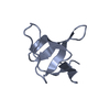 7pvqC  7pvsC 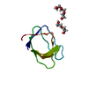 7pvvC 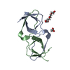 7pvwC 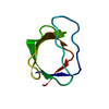 7pvyC  7pvzC  7pw0C 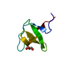 3eg3S S: Starting model for refinement C: citing same article ( |
|---|---|
| Similar structure data |
-Links
 PDBj
PDBj
- Assembly
| Deposited unit | 
| ||||||||
|---|---|---|---|---|---|---|---|---|---|
| 1 |
| ||||||||
| Unit cell |
|
-Components
| #1: Protein | Mass: 6721.397 Da / Num. of mol.: 1 / Mutation: V73E, A74S, S75R, G76T, D77E Source method: isolated from a genetically manipulated source Source: (gene. exp.) Homo sapiens (human) / Gene: BCR/ABL fusion / Plasmid: pHTP1 / Production host:  References: UniProt: A9UF07, non-specific protein-tyrosine kinase |
|---|---|
| #2: Water | ChemComp-HOH /  Mass: 18.015 Da / Num. of mol.: 51 / Source method: isolated from a natural source / Formula: H2O Mass: 18.015 Da / Num. of mol.: 51 / Source method: isolated from a natural source / Formula: H2O |
| Has protein modification | N |
-Experimental details
-Experiment
| Experiment | Method: X-RAY DIFFRACTION / Number of used crystals: 1 |
|---|
- Sample preparation
| Crystal | Density Matthews: 2.05 Å3/Da / Density % sol: 40.02 % / Mosaicity: 0.09 ° |
|---|---|
| Crystal grow | Temperature: 298 K / Method: vapor diffusion, sitting drop / pH: 7 / Details: 2.0 M ammonium sulfate, 0.1M HEPES |
-Data collection
| Diffraction | Mean temperature: 100 K / Serial crystal experiment: N | ||||||||||||||||||||||||||||||
|---|---|---|---|---|---|---|---|---|---|---|---|---|---|---|---|---|---|---|---|---|---|---|---|---|---|---|---|---|---|---|---|
| Diffraction source | Source: SYNCHROTRON / Site: ALBA / Beamline: XALOC / Wavelength: 0.97924 Å | ||||||||||||||||||||||||||||||
| Detector | Type: DECTRIS PILATUS 6M / Detector: PIXEL / Date: Dec 2, 2017 | ||||||||||||||||||||||||||||||
| Radiation | Protocol: SINGLE WAVELENGTH / Monochromatic (M) / Laue (L): M / Scattering type: x-ray | ||||||||||||||||||||||||||||||
| Radiation wavelength | Wavelength: 0.97924 Å / Relative weight: 1 | ||||||||||||||||||||||||||||||
| Reflection | Resolution: 1.1→17.44 Å / Num. obs: 21193 / % possible obs: 95.3 % / Redundancy: 2 % / Biso Wilson estimate: 17.78 Å2 / CC1/2: 0.998 / Rmerge(I) obs: 0.027 / Rpim(I) all: 0.022 / Rrim(I) all: 0.035 / Net I/σ(I): 9.9 / Num. measured all: 43012 | ||||||||||||||||||||||||||||||
| Reflection shell | Diffraction-ID: 1
|
-Phasing
| Phasing | Method: molecular replacement |
|---|
- Processing
| Software |
| ||||||||||||||||||||||||||||||||||||||||||||||||||||||||||||||||||||||||||||||||||||||||||||||||||
|---|---|---|---|---|---|---|---|---|---|---|---|---|---|---|---|---|---|---|---|---|---|---|---|---|---|---|---|---|---|---|---|---|---|---|---|---|---|---|---|---|---|---|---|---|---|---|---|---|---|---|---|---|---|---|---|---|---|---|---|---|---|---|---|---|---|---|---|---|---|---|---|---|---|---|---|---|---|---|---|---|---|---|---|---|---|---|---|---|---|---|---|---|---|---|---|---|---|---|---|
| Refinement | Method to determine structure: MOLECULAR REPLACEMENT Starting model: 3EG3 Resolution: 1.1→17.44 Å / SU ML: 0.12 / Cross valid method: THROUGHOUT / σ(F): 0.32 / Phase error: 34.68 / Stereochemistry target values: ML
| ||||||||||||||||||||||||||||||||||||||||||||||||||||||||||||||||||||||||||||||||||||||||||||||||||
| Solvent computation | Shrinkage radii: 0.9 Å / VDW probe radii: 1.11 Å / Solvent model: FLAT BULK SOLVENT MODEL | ||||||||||||||||||||||||||||||||||||||||||||||||||||||||||||||||||||||||||||||||||||||||||||||||||
| Displacement parameters | Biso max: 64.6 Å2 / Biso mean: 27.5203 Å2 / Biso min: 14.31 Å2 | ||||||||||||||||||||||||||||||||||||||||||||||||||||||||||||||||||||||||||||||||||||||||||||||||||
| Refinement step | Cycle: final / Resolution: 1.1→17.44 Å
| ||||||||||||||||||||||||||||||||||||||||||||||||||||||||||||||||||||||||||||||||||||||||||||||||||
| LS refinement shell | Refine-ID: X-RAY DIFFRACTION / Rfactor Rfree error: 0 / Total num. of bins used: 13
|
