 Movie
Movie Controller


+ Open data

- Basic information
| Entry | Database: PDB / ID: 7pqn | |||||||||||||||||||||
|---|---|---|---|---|---|---|---|---|---|---|---|---|---|---|---|---|---|---|---|---|---|---|
| Title | Catalytic fragment of MASP-2 in complex with ecotin | |||||||||||||||||||||
 Components Components |
| |||||||||||||||||||||
 Keywords Keywords | IMMUNE SYSTEM / serine protease / complement / inhibitor | |||||||||||||||||||||
| Function / homology |  Function and homology information Function and homology informationmannan-binding lectin-associated serine protease-2 / complement component C4b binding / Ficolins bind to repetitive carbohydrate structures on the target cell surface / Lectin pathway of complement activation / complement activation, lectin pathway / Initial triggering of complement / complement activation, classical pathway / serine-type endopeptidase inhibitor activity / defense response / calcium-dependent protein binding ...mannan-binding lectin-associated serine protease-2 / complement component C4b binding / Ficolins bind to repetitive carbohydrate structures on the target cell surface / Lectin pathway of complement activation / complement activation, lectin pathway / Initial triggering of complement / complement activation, classical pathway / serine-type endopeptidase inhibitor activity / defense response / calcium-dependent protein binding / peptidase activity / outer membrane-bounded periplasmic space / serine-type endopeptidase activity / calcium ion binding / SARS-CoV-2 activates/modulates innate and adaptive immune responses / protein homodimerization activity / proteolysis / extracellular space / extracellular exosome / extracellular region / identical protein binding Similarity search - Function | |||||||||||||||||||||
| Biological species |   Homo sapiens (human) Homo sapiens (human) | |||||||||||||||||||||
| Method |  X-RAY DIFFRACTION / SYNCHROTRON / MOLECULAR REPLACEMENT / Resolution: 2.40001520992 Å X-RAY DIFFRACTION / SYNCHROTRON / MOLECULAR REPLACEMENT / Resolution: 2.40001520992 Å | |||||||||||||||||||||
 Authors Authors | Harmat, V. / Fodor, K. / Heja, D. | |||||||||||||||||||||
| Funding support |  Hungary, 6items Hungary, 6items
| |||||||||||||||||||||
 Citation Citation | Journal: J.Biol.Chem. / Year: 2022 Title: Synergy of protease-binding sites within the ecotin homodimer is crucial for inhibition of MASP enzymes and for blocking lectin pathway activation. Authors: Nagy, Z.A. / Heja, D. / Bencze, D. / Kiss, B. / Boros, E. / Szakacs, D. / Fodor, K. / Wilmanns, M. / Kocsis, A. / Dobo, J. / Gal, P. / Harmat, V. / Pal, G. #1: Journal: Acta Crystallogr., Sect. D: Biol. Crystallogr. / Year: 2012 Title: Towards automated crystallographic structure refinement with phenix.refine. Authors: Afonine, P.V. / Grosse-Kunstleve, R.W. / Echols, N. / Headd, J.J. / Moriarty, N.W. / Mustyakimov, M. / Terwilliger, T.C. / Urzhumtsev, A. / Zwart, P.H. / Adams, P.D. #2: Journal: Acta Crystallogr D Biol Crystallogr / Year: 2010 Title: PHENIX: a comprehensive Python-based system for macromolecular structure solution. Authors: Paul D Adams / Pavel V Afonine / Gábor Bunkóczi / Vincent B Chen / Ian W Davis / Nathaniel Echols / Jeffrey J Headd / Li-Wei Hung / Gary J Kapral / Ralf W Grosse-Kunstleve / Airlie J McCoy ...Authors: Paul D Adams / Pavel V Afonine / Gábor Bunkóczi / Vincent B Chen / Ian W Davis / Nathaniel Echols / Jeffrey J Headd / Li-Wei Hung / Gary J Kapral / Ralf W Grosse-Kunstleve / Airlie J McCoy / Nigel W Moriarty / Robert Oeffner / Randy J Read / David C Richardson / Jane S Richardson / Thomas C Terwilliger / Peter H Zwart /  Abstract: Macromolecular X-ray crystallography is routinely applied to understand biological processes at a molecular level. However, significant time and effort are still required to solve and complete many ...Macromolecular X-ray crystallography is routinely applied to understand biological processes at a molecular level. However, significant time and effort are still required to solve and complete many of these structures because of the need for manual interpretation of complex numerical data using many software packages and the repeated use of interactive three-dimensional graphics. PHENIX has been developed to provide a comprehensive system for macromolecular crystallographic structure solution with an emphasis on the automation of all procedures. This has relied on the development of algorithms that minimize or eliminate subjective input, the development of algorithms that automate procedures that are traditionally performed by hand and, finally, the development of a framework that allows a tight integration between the algorithms. #4: Journal: Acta Crystallogr D Biol Crystallogr / Year: 2010 Title: Molecular replacement with MOLREP. Authors: Vagin, A. / Teplyakov, A. #5: Journal: PLoS Pathog / Year: 2019Title: Ecotin, a microbial inhibitor of serine proteases, blocks multiple complement dependent and independent microbicidal activities of human serum. Authors: Nagy, Z.A. / Szakacs, D. / Boros, E. / Heja, D. / Vigh, E. / Sandor, N. / Jozsi, M. / Oroszlan, G. / Dobo, J. / Gal, P. / Pal, G. | |||||||||||||||||||||
| History |
|
- Structure visualization
| Structure viewer | Molecule: MolmilJmol/JSmol |
|---|
- Downloads & links
-Download
| PDBx/mmCIF format | 7pqn.cif.gz | 428.3 KB | Display | PDBx/mmCIF format |
|---|---|---|---|---|
| PDB format | pdb7pqn.ent.gz | Display | PDB format | |
| PDBx/mmJSON format | 7pqn.json.gz | Tree view | PDBx/mmJSON format | |
| Others |  Other downloads Other downloads |
-Validation report
| Arichive directory | https://data.pdbj.org/pub/pdb/validation_reports/pq/7pqnftp://data.pdbj.org/pub/pdb/validation_reports/pq/7pqn | HTTPS FTP |
|---|
-Related structure data
| Related structure data |  7pqoC  1azzS  1q3xS S: Starting model for refinement C: citing same article ( |
|---|---|
| Similar structure data |
-Links
 PDBj
PDBj


- Assembly
| Deposited unit | 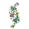
| |||||||||||||||||||||||||||||||||||||||||||||||||||||||||||||||||||||||||||||||||||||||||||||||||||
|---|---|---|---|---|---|---|---|---|---|---|---|---|---|---|---|---|---|---|---|---|---|---|---|---|---|---|---|---|---|---|---|---|---|---|---|---|---|---|---|---|---|---|---|---|---|---|---|---|---|---|---|---|---|---|---|---|---|---|---|---|---|---|---|---|---|---|---|---|---|---|---|---|---|---|---|---|---|---|---|---|---|---|---|---|---|---|---|---|---|---|---|---|---|---|---|---|---|---|---|---|
| 1 |
| |||||||||||||||||||||||||||||||||||||||||||||||||||||||||||||||||||||||||||||||||||||||||||||||||||
| Unit cell |
| |||||||||||||||||||||||||||||||||||||||||||||||||||||||||||||||||||||||||||||||||||||||||||||||||||
| Noncrystallographic symmetry (NCS) | NCS domain:
NCS domain segments:
|