 Movie
Movie Controller

[English] 日本語
 Yorodumi
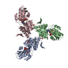
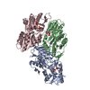
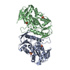
- PDB-7pn0: Crystal structure of the Phosphorybosylpyrophosphate synthetase I... -
+ Open data

- Basic information
| Entry | Database: PDB / ID: 7pn0 | |||||||||
|---|---|---|---|---|---|---|---|---|---|---|
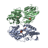
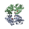
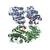
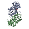
| Title | Crystal structure of the Phosphorybosylpyrophosphate synthetase II from Thermus thermophilus at R32 space group | |||||||||
 Components Components | Ribose-phosphate pyrophosphokinase | |||||||||
 Keywords Keywords | TRANSFERASE | |||||||||
| Function / homology |  Function and homology information Function and homology informationribonucleoside monophosphate biosynthetic process / ribose-phosphate diphosphokinase / ribose phosphate diphosphokinase activity / nucleotide biosynthetic process / 5-phosphoribose 1-diphosphate biosynthetic process / nucleoside metabolic process / kinase activity / magnesium ion binding / ATP binding / cytoplasm Similarity search - Function | |||||||||
| Biological species |   Thermus thermophilus (bacteria) Thermus thermophilus (bacteria) | |||||||||
| Method |  X-RAY DIFFRACTION / SYNCHROTRON / MOLECULAR REPLACEMENT / Resolution: 1.85 Å X-RAY DIFFRACTION / SYNCHROTRON / MOLECULAR REPLACEMENT / Resolution: 1.85 Å | |||||||||
 Authors Authors | Timofeev, V.I. / Abramchik, Y.A. / Kostromina, M.A. / Esipov, R.S. / Kuranova, I.P. | |||||||||
| Funding support | 1items
| |||||||||
 Citation Citation | Journal: To Be Published Title: Crystal structure of the Phosphorybosylpyrophosphate synthetase II from Thermus thermophilus at R32 space group Authors: Timofeev, V.I. / Abramchik, Y.A. / Kostromina, M.A. / Esipov, R.S. / Kuranova, I.P. | |||||||||
| History |
|
- Structure visualization
| Structure viewer | Molecule: MolmilJmol/JSmol |
|---|
- Downloads & links
-Download
| PDBx/mmCIF format | 7pn0.cif.gz | 137.5 KB | Display | PDBx/mmCIF format |
|---|---|---|---|---|
| PDB format | pdb7pn0.ent.gz | 106.7 KB | Display | PDB format |
| PDBx/mmJSON format | 7pn0.json.gz | Tree view | PDBx/mmJSON format | |
| Others |  Other downloads Other downloads |
-Validation report
| Arichive directory | https://data.pdbj.org/pub/pdb/validation_reports/pn/7pn0ftp://data.pdbj.org/pub/pdb/validation_reports/pn/7pn0 | HTTPS FTP |
|---|
-Related structure data
| Related structure data |  5t3oS S: Starting model for refinement |
|---|---|
| Similar structure data |

-Links
 PDBj
PDBj
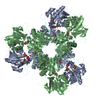
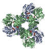
- Assembly
| Deposited unit | 
| ||||||||
|---|---|---|---|---|---|---|---|---|---|
| 1 | 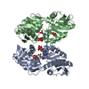
| ||||||||
| Unit cell |
|
-Components
| #1: Protein | Mass: 34364.445 Da / Num. of mol.: 2 Source method: isolated from a genetically manipulated source Source: (gene. exp.) Thermus thermophilus (bacteria) / Gene: prs, HB27c_C1311 / Production host: Thermus thermophilus (bacteria)References: UniProt: A0A7G5B4V6, ribose-phosphate diphosphokinase #2: Chemical |   Mass: 427.201 Da / Num. of mol.: 2 / Source method: obtained synthetically / Formula: C10H15N5O10P2 / Comment: ADP, energy-carrying molecule*YM Mass: 427.201 Da / Num. of mol.: 2 / Source method: obtained synthetically / Formula: C10H15N5O10P2 / Comment: ADP, energy-carrying molecule*YM#3: Chemical | ChemComp-SO4 /   Mass: 96.063 Da / Num. of mol.: 8 / Source method: obtained synthetically / Formula: SO4 Mass: 96.063 Da / Num. of mol.: 8 / Source method: obtained synthetically / Formula: SO4#4: Water | ChemComp-HOH / |  Mass: 18.015 Da / Num. of mol.: 252 / Source method: isolated from a natural source / Formula: H2O Mass: 18.015 Da / Num. of mol.: 252 / Source method: isolated from a natural source / Formula: H2OHas ligand of interest | N | |
|---|
-Experimental details
-Experiment
| Experiment | Method: X-RAY DIFFRACTION / Number of used crystals: 1 |
|---|
- Sample preparation
| Crystal | Density Matthews: 2.7 Å3/Da / Density % sol: 54.44 % |
|---|---|
| Crystal grow | Temperature: 293 K / Method: counter-diffusion Details: 1,7M lithium sulphate, 0,1M HEPES pH 7.5, 0,04%NaN3, 5mM MgCl2 |
-Data collection
| Diffraction | Mean temperature: 100 K / Serial crystal experiment: N |
|---|---|
| Diffraction source | Source: SYNCHROTRON / Site: SPring-8  / Beamline: BL41XU / Wavelength: 0.979 Å / Beamline: BL41XU / Wavelength: 0.979 Å |
| Detector | Type: DECTRIS EIGER X 16M / Detector: PIXEL / Date: Oct 10, 2019 |
| Radiation | Protocol: SINGLE WAVELENGTH / Monochromatic (M) / Laue (L): M / Scattering type: x-ray |
| Radiation wavelength | Wavelength: 0.979 Å / Relative weight: 1 |
| Reflection | Resolution: 1.85→20 Å / Num. obs: 59412 / % possible obs: 100 % / Redundancy: 7.01 % / CC1/2: 0.999 / Rmerge(I) obs: 0.105 / Net I/σ(I): 14.31 |
| Reflection shell | Resolution: 1.85→1.89 Å / Rmerge(I) obs: 0.546 / Num. unique obs: 4323 / CC1/2: 0.905 |
- Processing
| Software |
| ||||||||||||||||||||||||||||||||||||||||||||||||||||||||||||
|---|---|---|---|---|---|---|---|---|---|---|---|---|---|---|---|---|---|---|---|---|---|---|---|---|---|---|---|---|---|---|---|---|---|---|---|---|---|---|---|---|---|---|---|---|---|---|---|---|---|---|---|---|---|---|---|---|---|---|---|---|---|
| Refinement | Method to determine structure: MOLECULAR REPLACEMENT Starting model: 5T3O Resolution: 1.85→19.77 Å / Cor.coef. Fo:Fc: 0.953 / Cor.coef. Fo:Fc free: 0.93 / SU B: 2.92 / SU ML: 0.087 / Cross valid method: THROUGHOUT / σ(F): 0 / ESU R: 0.127 / ESU R Free: 0.122 / Stereochemistry target values: MAXIMUM LIKELIHOOD Details: HYDROGENS HAVE BEEN ADDED IN THE RIDING POSITIONS U VALUES : REFINED INDIVIDUALLY
| ||||||||||||||||||||||||||||||||||||||||||||||||||||||||||||
| Solvent computation | Ion probe radii: 0.8 Å / Shrinkage radii: 0.8 Å / VDW probe radii: 1.2 Å / Solvent model: MASK | ||||||||||||||||||||||||||||||||||||||||||||||||||||||||||||
| Displacement parameters | Biso max: 105.84 Å2 / Biso mean: 23.235 Å2 / Biso min: 9.7 Å2
| ||||||||||||||||||||||||||||||||||||||||||||||||||||||||||||
| Refinement step | Cycle: final / Resolution: 1.85→19.77 Å
| ||||||||||||||||||||||||||||||||||||||||||||||||||||||||||||
| Refine LS restraints |
| ||||||||||||||||||||||||||||||||||||||||||||||||||||||||||||
| LS refinement shell | Resolution: 1.85→1.898 Å / Rfactor Rfree error: 0 / Total num. of bins used: 20
|
