 Movie
Movie Controller

[English] 日本語
 Yorodumi
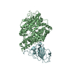
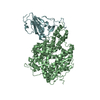
- PDB-7pki: Crystal structure of human ACE2 bound to the spike receptor-bindi... -
+ Open data

- Basic information
| Entry | Database: PDB / ID: 7pki | ||||||
|---|---|---|---|---|---|---|---|
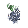
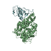
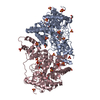
| Title | Crystal structure of human ACE2 bound to the spike receptor-binding domain from a cave bat sarbecovirus closely related to SARS-CoV-2. | ||||||
 Components Components |
| ||||||
 Keywords Keywords | VIRAL PROTEIN / Coronavirus / receptor binding domain / human ACE2. | ||||||
| Function / homology |  Function and homology information Function and homology informationpositive regulation of amino acid transport / angiotensin-converting enzyme 2 / positive regulation of L-proline import across plasma membrane / Hydrolases; Acting on peptide bonds (peptidases); Metallocarboxypeptidases / angiotensin-mediated drinking behavior / positive regulation of gap junction assembly / regulation of systemic arterial blood pressure by renin-angiotensin / tryptophan transport / regulation of cardiac conduction / maternal process involved in female pregnancy ...positive regulation of amino acid transport / angiotensin-converting enzyme 2 / positive regulation of L-proline import across plasma membrane / Hydrolases; Acting on peptide bonds (peptidases); Metallocarboxypeptidases / angiotensin-mediated drinking behavior / positive regulation of gap junction assembly / regulation of systemic arterial blood pressure by renin-angiotensin / tryptophan transport / regulation of cardiac conduction / maternal process involved in female pregnancy / peptidyl-dipeptidase activity / regulation of vasoconstriction / transporter activator activity / Metabolism of Angiotensinogen to Angiotensins / carboxypeptidase activity / angiotensin maturation / viral life cycle / Attachment and Entry / receptor-mediated endocytosis of virus by host cell / metallocarboxypeptidase activity / positive regulation of cardiac muscle contraction / regulation of cytokine production / blood vessel diameter maintenance / negative regulation of smooth muscle cell proliferation / brush border membrane / negative regulation of ERK1 and ERK2 cascade / positive regulation of reactive oxygen species metabolic process / metallopeptidase activity / endocytic vesicle membrane / regulation of cell population proliferation / virus receptor activity / regulation of inflammatory response / endopeptidase activity / viral translation / Induction of Cell-Cell Fusion / Potential therapeutics for SARS / membrane fusion / entry receptor-mediated virion attachment to host cell / Attachment and Entry / receptor-mediated virion attachment to host cell / cilium / apical plasma membrane / membrane raft / endoplasmic reticulum lumen / symbiont entry into host cell / cell surface / negative regulation of transcription by RNA polymerase II / extracellular space / extracellular exosome / extracellular region / zinc ion binding / identical protein binding / membrane / plasma membrane Similarity search - Function | ||||||
| Biological species |  Homo sapiens (human) Homo sapiens (human) Sarbecovirus Sarbecovirus | ||||||
| Method |  X-RAY DIFFRACTION / SYNCHROTRON / MOLECULAR REPLACEMENT / Resolution: 2.94234133444 Å X-RAY DIFFRACTION / SYNCHROTRON / MOLECULAR REPLACEMENT / Resolution: 2.94234133444 Å | ||||||
 Authors Authors | Baquero, E. / Rey, F.A. | ||||||
| Funding support |  France, 1items France, 1items
| ||||||
 Citation Citation | Journal: Nature / Year: 2022 Title: Bat coronaviruses related to SARS-CoV-2 and infectious for human cells. Authors: Temmam, S. / Vongphayloth, K. / Baquero, E. / Munier, S. / Bonomi, M. / Regnault, B. / Douangboubpha, B. / Karami, Y. / Chretien, D. / Sanamxay, D. / Xayaphet, V. / Paphaphanh, P. / Lacoste, ...Authors: Temmam, S. / Vongphayloth, K. / Baquero, E. / Munier, S. / Bonomi, M. / Regnault, B. / Douangboubpha, B. / Karami, Y. / Chretien, D. / Sanamxay, D. / Xayaphet, V. / Paphaphanh, P. / Lacoste, V. / Somlor, S. / Lakeomany, K. / Phommavanh, N. / Perot, P. / Dehan, O. / Amara, F. / Donati, F. / Bigot, T. / Nilges, M. / Rey, F.A. / van der Werf, S. / Brey, P.T. / Eloit, M. | ||||||
| History |
|
- Structure visualization
| Structure viewer | Molecule: MolmilJmol/JSmol |
|---|
- Downloads & links
-Download
| PDBx/mmCIF format | 7pki.cif.gz | 221.1 KB | Display | PDBx/mmCIF format |
|---|---|---|---|---|
| PDB format | pdb7pki.ent.gz | 139.9 KB | Display | PDB format |
| PDBx/mmJSON format | 7pki.json.gz | Tree view | PDBx/mmJSON format | |
| Others |  Other downloads Other downloads |
-Validation report
| Summary document | 7pki_validation.pdf.gz | 835.2 KB | Display | wwPDB validaton report |
|---|---|---|---|---|
| Full document | 7pki_full_validation.pdf.gz | 840.5 KB | Display | |
| Data in XML | 7pki_validation.xml.gz | 29.3 KB | Display | |
| Data in CIF | 7pki_validation.cif.gz | 39.4 KB | Display | |
| Arichive directory | https://data.pdbj.org/pub/pdb/validation_reports/pk/7pkiftp://data.pdbj.org/pub/pdb/validation_reports/pk/7pki | HTTPS FTP |
-Related structure data
| Related structure data | 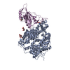 6m0jS S: Starting model for refinement |
|---|---|
| Similar structure data |


-Links
 PDBj
PDBj


- Assembly
| Deposited unit | 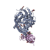
| ||||||||||||
|---|---|---|---|---|---|---|---|---|---|---|---|---|---|
| 1 |
| ||||||||||||
| Unit cell |
| ||||||||||||
| Components on special symmetry positions |
|
-Components
-Protein , 2 types, 2 molecules AE
| #1: Protein | Mass: 69038.578 Da / Num. of mol.: 1 Source method: isolated from a genetically manipulated source Source: (gene. exp.) Homo sapiens (human) / Gene: ACE2, UNQ868/PRO1885 / Plasmid: pcDNA3.1(+) / Cell line (production host): Expi293 GnTI / Production host: Homo sapiens (human) / References: UniProt: Q9BYF1 |
|---|---|
| #2: Protein | Mass: 22062.768 Da / Num. of mol.: 1 Source method: isolated from a genetically manipulated source Source: (gene. exp.) Sarbecovirus / Strain: BANAL 236 / Gene: Spike / Plasmid: pCAGGS / Cell line (production host): Espi293 GnTI / Production host: Homo sapiens (human) |
-Sugars , 2 types, 7 molecules 
| #3: Polysaccharide | 2-acetamido-2-deoxy-beta-D-glucopyranose-(1-4)-2-acetamido-2-deoxy-beta-D-glucopyranose Source method: isolated from a genetically manipulated source |
|---|---|
| #4: Sugar | ChemComp-NAG /  Type: D-saccharide, beta linking / Mass: 221.208 Da / Num. of mol.: 6 / Source method: obtained synthetically / Formula: C8H15NO6 Type: D-saccharide, beta linking / Mass: 221.208 Da / Num. of mol.: 6 / Source method: obtained synthetically / Formula: C8H15NO6 |
-Non-polymers , 7 types, 22 molecules 


| #5: Chemical | ChemComp-SO4 /  Mass: 96.063 Da / Num. of mol.: 4 / Source method: obtained synthetically / Formula: SO4 Mass: 96.063 Da / Num. of mol.: 4 / Source method: obtained synthetically / Formula: SO4#6: Chemical | ChemComp-TRS / |  Mass: 122.143 Da / Num. of mol.: 1 / Source method: obtained synthetically / Formula: C4H12NO3 / Comment: pH buffer*YM Mass: 122.143 Da / Num. of mol.: 1 / Source method: obtained synthetically / Formula: C4H12NO3 / Comment: pH buffer*YM#7: Chemical | ChemComp-PEG /  Mass: 106.120 Da / Num. of mol.: 5 / Source method: obtained synthetically / Formula: C4H10O3 Mass: 106.120 Da / Num. of mol.: 5 / Source method: obtained synthetically / Formula: C4H10O3#8: Chemical | ChemComp-ZN / |  Mass: 65.409 Da / Num. of mol.: 1 / Source method: isolated from a natural source / Formula: Zn Mass: 65.409 Da / Num. of mol.: 1 / Source method: isolated from a natural source / Formula: Zn#9: Chemical |  Mass: 35.453 Da / Num. of mol.: 2 / Source method: obtained synthetically / Formula: Cl Mass: 35.453 Da / Num. of mol.: 2 / Source method: obtained synthetically / Formula: Cl#10: Chemical | ChemComp-GOL / |  Mass: 92.094 Da / Num. of mol.: 1 / Source method: obtained synthetically / Formula: C3H8O3 Mass: 92.094 Da / Num. of mol.: 1 / Source method: obtained synthetically / Formula: C3H8O3#11: Water | ChemComp-HOH / | Mass: 18.015 Da / Num. of mol.: 8 / Source method: isolated from a natural source / Formula: H2O |
|---|
-Details
| Has ligand of interest | N |
|---|---|
| Has protein modification | Y |
-Experimental details
-Experiment
| Experiment | Method: X-RAY DIFFRACTION / Number of used crystals: 1 |
|---|
- Sample preparation
| Crystal | Density Matthews: 6.17 Å3/Da / Density % sol: 80 % |
|---|---|
| Crystal grow | Temperature: 277.15 K / Method: vapor diffusion, sitting drop / pH: 8.5 Details: 0.2 M Lithium sulfate, 0.1 M Tris 8.5, 30 % w/v PEG 4000 |
-Data collection
| Diffraction | Mean temperature: 80 K / Serial crystal experiment: N |
|---|---|
| Diffraction source | Source: SYNCHROTRON / Site: SOLEIL / Beamline: PROXIMA 1 / Wavelength: 0.978 Å |
| Detector | Type: DECTRIS EIGER X 16M / Detector: PIXEL / Date: Jul 27, 2021 |
| Radiation | Protocol: SINGLE WAVELENGTH / Monochromatic (M) / Laue (L): M / Scattering type: x-ray |
| Radiation wavelength | Wavelength: 0.978 Å / Relative weight: 1 |
| Reflection | Resolution: 2.942→49.63 Å / Num. obs: 43645 / % possible obs: 86.98 % / Redundancy: 2 % / Biso Wilson estimate: 68.4 Å2 / CC1/2: 0.996 / Rmerge(I) obs: 0.05 / Net I/σ(I): 11.42 |
| Reflection shell | Resolution: 2.942→3.047 Å / Rmerge(I) obs: 4.562 / Num. unique obs: 614 / CC1/2: 0.496 |
- Processing
| Software |
| ||||||||||||||||||||||||||||||||||||||||||||||||||||||||||||||||||||||||||||||||||||||||||||||||||||||||||||||||
|---|---|---|---|---|---|---|---|---|---|---|---|---|---|---|---|---|---|---|---|---|---|---|---|---|---|---|---|---|---|---|---|---|---|---|---|---|---|---|---|---|---|---|---|---|---|---|---|---|---|---|---|---|---|---|---|---|---|---|---|---|---|---|---|---|---|---|---|---|---|---|---|---|---|---|---|---|---|---|---|---|---|---|---|---|---|---|---|---|---|---|---|---|---|---|---|---|---|---|---|---|---|---|---|---|---|---|---|---|---|---|---|---|---|
| Refinement | Method to determine structure: MOLECULAR REPLACEMENT Starting model: 6M0J Resolution: 2.94234133444→49.2963310469 Å / SU ML: 0.293331497159 / Cross valid method: FREE R-VALUE / σ(F): 1.34352455118 / Phase error: 25.2228022368 Stereochemistry target values: GeoStd + Monomer Library + CDL v1.2
| ||||||||||||||||||||||||||||||||||||||||||||||||||||||||||||||||||||||||||||||||||||||||||||||||||||||||||||||||
| Solvent computation | Shrinkage radii: 0.9 Å / VDW probe radii: 1.11 Å / Solvent model: FLAT BULK SOLVENT MODEL | ||||||||||||||||||||||||||||||||||||||||||||||||||||||||||||||||||||||||||||||||||||||||||||||||||||||||||||||||
| Displacement parameters | Biso mean: 64.6809885402 Å2 | ||||||||||||||||||||||||||||||||||||||||||||||||||||||||||||||||||||||||||||||||||||||||||||||||||||||||||||||||
| Refinement step | Cycle: LAST / Resolution: 2.94234133444→49.2963310469 Å
| ||||||||||||||||||||||||||||||||||||||||||||||||||||||||||||||||||||||||||||||||||||||||||||||||||||||||||||||||
| Refine LS restraints |
| ||||||||||||||||||||||||||||||||||||||||||||||||||||||||||||||||||||||||||||||||||||||||||||||||||||||||||||||||
| LS refinement shell |
|
