[English] 日本語
Yorodumi
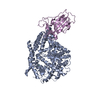
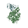
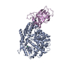
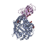
- PDB-3scl: Crystal structure of spike protein receptor-binding domain from S... -

+
Open data


ID or keywords:

Loading...

-
Basic information

Entry
Database: PDB / ID: 3scl
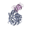
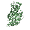
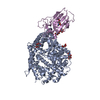
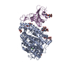
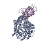
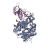
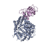
TitleCrystal structure of spike protein receptor-binding domain from SARS coronavirus epidemic strain complexed with human-civet chimeric receptor ACE2
Components
  • Angiotensin-converting enzyme 2 chimera
  • Spike glycoprotein
KeywordsHYDROLASE/VIRAL PROTEIN / beta-sheet / HYDROLASE-VIRAL PROTEIN complex
Function / homology
Function and homology information


Maturation of spike protein / Translation of Structural Proteins / Virion Assembly and Release / positive regulation of amino acid transport / angiotensin-converting enzyme 2 / positive regulation of L-proline import across plasma membrane / Hydrolases; Acting on peptide bonds (peptidases); Metallocarboxypeptidases / angiotensin-mediated drinking behavior / regulation of systemic arterial blood pressure by renin-angiotensin / tryptophan transport ...Maturation of spike protein / Translation of Structural Proteins / Virion Assembly and Release / positive regulation of amino acid transport / angiotensin-converting enzyme 2 / positive regulation of L-proline import across plasma membrane / Hydrolases; Acting on peptide bonds (peptidases); Metallocarboxypeptidases / angiotensin-mediated drinking behavior / regulation of systemic arterial blood pressure by renin-angiotensin / tryptophan transport / positive regulation of gap junction assembly / regulation of cardiac conduction / regulation of vasoconstriction / peptidyl-dipeptidase activity / maternal process involved in female pregnancy / angiotensin maturation / Metabolism of Angiotensinogen to Angiotensins / negative regulation of signaling receptor activity / carboxypeptidase activity / Attachment and Entry / positive regulation of cardiac muscle contraction / viral life cycle / regulation of cytokine production / blood vessel diameter maintenance / negative regulation of smooth muscle cell proliferation / brush border membrane / regulation of transmembrane transporter activity / endocytosis involved in viral entry into host cell / cilium / negative regulation of ERK1 and ERK2 cascade / SARS-CoV-1 activates/modulates innate immune responses / metallopeptidase activity / positive regulation of reactive oxygen species metabolic process / endocytic vesicle membrane / virus receptor activity / regulation of cell population proliferation / regulation of inflammatory response / endopeptidase activity / suppression by virus of host tetherin activity / Potential therapeutics for SARS / Induction of Cell-Cell Fusion / entry receptor-mediated virion attachment to host cell / host cell endoplasmic reticulum-Golgi intermediate compartment membrane / membrane fusion / receptor-mediated endocytosis of virus by host cell / Attachment and Entry / positive regulation of viral entry into host cell / receptor-mediated virion attachment to host cell / host cell surface receptor binding / symbiont-mediated suppression of host innate immune response / symbiont entry into host cell / membrane raft / apical plasma membrane / endoplasmic reticulum lumen / fusion of virus membrane with host plasma membrane / fusion of virus membrane with host endosome membrane / viral envelope / host cell plasma membrane / virion membrane / cell surface / proteolysis / extracellular space / extracellular exosome / zinc ion binding / extracellular region / identical protein binding / membrane / metal ion binding / plasma membrane / cytoplasm
Similarity search - Function
Spike protein, C-terminal core receptor binding subdomain / Spike (S) protein S1 subunit, receptor-binding domain, SARS-CoV / Collectrin domain / Renal amino acid transporter / Collectrin-like domain profile. / Peptidase M2, peptidyl-dipeptidase A / Angiotensin-converting enzyme / Peptidase family M2 domain profile. / Neutral zinc metallopeptidases, zinc-binding region signature. / Spike (S) protein S1 subunit, N-terminal domain, SARS-CoV-like ...Spike protein, C-terminal core receptor binding subdomain / Spike (S) protein S1 subunit, receptor-binding domain, SARS-CoV / Collectrin domain / Renal amino acid transporter / Collectrin-like domain profile. / Peptidase M2, peptidyl-dipeptidase A / Angiotensin-converting enzyme / Peptidase family M2 domain profile. / Neutral zinc metallopeptidases, zinc-binding region signature. / Spike (S) protein S1 subunit, N-terminal domain, SARS-CoV-like / Spike glycoprotein, betacoronavirus / Spike glycoprotein, N-terminal domain superfamily / Betacoronavirus spike (S) glycoprotein S1 subunit N-terminal (NTD) domain profile. / Betacoronavirus spike (S) glycoprotein S1 subunit C-terminal (CTD) domain profile. / Spike (S) protein S1 subunit, receptor-binding domain, betacoronavirus / Spike S1 subunit, receptor binding domain superfamily, betacoronavirus / Spike glycoprotein S1, N-terminal domain, betacoronavirus-like / Betacoronavirus-like spike glycoprotein S1, N-terminal / Betacoronavirus spike glycoprotein S1, receptor binding / Spike glycoprotein S2, coronavirus, heptad repeat 1 / Spike glycoprotein S2, coronavirus, heptad repeat 2 / Coronavirus spike (S) glycoprotein S2 subunit heptad repeat 1 (HR1) region profile. / Coronavirus spike (S) glycoprotein S2 subunit heptad repeat 2 (HR2) region profile. / Spike glycoprotein S2 superfamily, coronavirus / Spike glycoprotein S2, coronavirus / Coronavirus spike glycoprotein S2 / Alpha-Beta Plaits / 2-Layer Sandwich / Alpha Beta
Similarity search - Domain/homology
Spike glycoprotein / Angiotensin-converting enzyme 2 / Angiotensin-converting enzyme 2
Similarity search - Component
Biological speciesPaguma larvata (masked palm civet)
Homo sapiens (human)
SARS coronavirus
MethodX-RAY DIFFRACTION / SYNCHROTRON / MOLECULAR REPLACEMENT / Resolution: 3 Å
AuthorsWu, K. / Peng, G. / Wilken, M. / Geraghty, R. / Li, F.
CitationJournal: J.Biol.Chem. / Year: 2012
Title: Mechanisms of host receptor adaptation by severe acute respiratory syndrome coronavirus.
Authors: Wu, K. / Peng, G. / Wilken, M. / Geraghty, R.J. / Li, F.
History
DepositionJun 7, 2011Deposition site: RCSB / Processing site: RCSB
Revision 1.0Feb 8, 2012Provider: repository / Type: Initial release
Revision 1.1Aug 8, 2012Group: Data collection
Revision 1.2Jul 26, 2017Group: Refinement description / Source and taxonomy / Category: entity_src_gen / software / Item: _software.classification
Revision 1.3Jul 17, 2019Group: Data collection / Refinement description / Category: software
Item: _software.classification / _software.name / _software.version
Revision 1.4Sep 16, 2020Group: Database references / Derived calculations
Category: citation / citation_author ...citation / citation_author / pdbx_struct_conn_angle / struct_conn / struct_ref_seq_dif / struct_site
Item: _citation.country / _citation.journal_abbrev ..._citation.country / _citation.journal_abbrev / _citation.journal_id_ASTM / _citation.journal_id_CSD / _citation.journal_id_ISSN / _citation.journal_volume / _citation.page_first / _citation.page_last / _citation.pdbx_database_id_DOI / _citation.pdbx_database_id_PubMed / _citation.title / _citation.year / _citation_author.name / _pdbx_struct_conn_angle.ptnr1_auth_seq_id / _pdbx_struct_conn_angle.ptnr1_label_seq_id / _pdbx_struct_conn_angle.ptnr3_auth_seq_id / _pdbx_struct_conn_angle.ptnr3_label_seq_id / _pdbx_struct_conn_angle.value / _struct_conn.pdbx_dist_value / _struct_conn.ptnr1_auth_asym_id / _struct_conn.ptnr1_auth_seq_id / _struct_conn.ptnr1_label_asym_id / _struct_conn.ptnr1_label_seq_id / _struct_conn.ptnr2_auth_asym_id / _struct_conn.ptnr2_label_asym_id / _struct_ref_seq_dif.details / _struct_site.pdbx_auth_asym_id / _struct_site.pdbx_auth_comp_id / _struct_site.pdbx_auth_seq_id

-
Structure visualization

Structure viewerMolecule:
MolmilJmol/JSmol

Downloads & links

-
Assembly

Deposited unit
A: Angiotensin-converting enzyme 2 chimera
B: Angiotensin-converting enzyme 2 chimera
E: Spike glycoprotein
F: Spike glycoprotein
hetero molecules


Theoretical massNumber of molelcules
Total (without water)182,4508
Polymers182,2484
Non-polymers2024
Water00
1
A: Angiotensin-converting enzyme 2 chimera
E: Spike glycoprotein
hetero molecules


Theoretical massNumber of molelcules
Total (without water)91,2254
Polymers91,1242
Non-polymers1012
Water0
TypeNameSymmetry operationNumber
identity operation1_555x,y,z1
Buried area2100 Å2
ΔGint-49 kcal/mol
Surface area33920 Å2
MethodPISA
2
B: Angiotensin-converting enzyme 2 chimera
F: Spike glycoprotein
hetero molecules


Theoretical massNumber of molelcules
Total (without water)91,2254
Polymers91,1242
Non-polymers1012
Water0
TypeNameSymmetry operationNumber
identity operation1_555x,y,z1
Unit cell
Length a, b, c (Å)81.670, 119.523, 113.532
Angle α, β, γ (deg.)90.00, 92.50, 90.00
Int Tables number4
Space group name H-MP1211
Noncrystallographic symmetry (NCS)NCS domain:
IDEns-IDDetails
11A
21B
12A
22B
13E
23F

NCS domain segments:
Dom-IDComponent-IDEns-IDRefine codeAuth asym-IDAuth seq-ID
1116A19 - 102
2116B19 - 102
1216A290 - 397
2216B290 - 397
1316A417 - 430
2316B417 - 430
1126A103 - 289
2126B103 - 289
1226A398 - 416
2226B398 - 416
1326A431 - 615
2326B431 - 615
1136E323 - 506
2136F323 - 506

NCS ensembles :
ID
1
2
3

-
Components

#1: Protein Angiotensin-converting enzyme 2 chimera / ACE2 / ACE-related carboxypeptidase / Angiotensin-converting enzyme homolog / ACEH / ...ACE2 / ACE-related carboxypeptidase / Angiotensin-converting enzyme homolog / ACEH / Metalloprotease MPROT15 / Processed angiotensin-converting enzyme 2


Mass: 69875.344 Da / Num. of mol.: 2 / Fragment: SEE REMARK 999
Source method: isolated from a genetically manipulated source
Source: (gene. exp.) Paguma larvata (masked palm civet), (gene. exp.) Homo sapiens (human)
Gene: ACE2, UNQ868/PRO1885 / Production host: Spodoptera frugiperda (fall armyworm)
References: UniProt: Q56NL1, UniProt: Q9BYF1, angiotensin-converting enzyme 2
#2: Protein Spike glycoprotein / S glycoprotein / E2 / Peplomer protein / Spike protein S1


Mass: 21248.811 Da / Num. of mol.: 2 / Fragment: receptor binding domain (UNP residues 324-502)
Source method: isolated from a genetically manipulated source
Source: (gene. exp.) SARS coronavirus / Gene: S, 2 / Production host: Spodoptera frugiperda (fall armyworm) / References: UniProt: P59594
#3: Chemical ChemComp-ZN / ZINC ION


Mass: 65.409 Da / Num. of mol.: 2 / Source method: obtained synthetically / Formula: Zn
#4: Chemical ChemComp-CL / CHLORIDE ION


Mass: 35.453 Da / Num. of mol.: 2 / Source method: obtained synthetically / Formula: Cl
Sequence detailsCHAINS A AND B ARE CHIMERIC, COMPRISING RESIDUES 19-82 OF UNP Q56NL1 AND RESIDUES 83-615 OF UNP Q9BYF1.

-
Experimental details

-
Experiment

ExperimentMethod: X-RAY DIFFRACTION / Number of used crystals: 1

-
Sample preparation

CrystalDensity Matthews: 3.04 Å3/Da / Density % sol: 59.51 %
Crystal growTemperature: 295 K / Method: evaporation / pH: 8.5
Details: 100 mM Tris, pH 8.5, 20% PEG6000, 100 mM sodium chloride, EVAPORATION, temperature 295K

-
Data collection

DiffractionMean temperature: 100 K
Diffraction sourceSource: SYNCHROTRON / Site: APS / Beamline: 24-ID-E
DetectorType: ADSC QUANTUM 315 / Detector: CCD
RadiationMonochromator: single crystal Si(220) side bounce / Protocol: SINGLE WAVELENGTH / Monochromatic (M) / Laue (L): M / Scattering type: x-ray
Radiation wavelengthRelative weight: 1
ReflectionResolution: 3→50 Å / Num. all: 43744 / Num. obs: 42738 / % possible obs: 97.7 % / Observed criterion σ(F): 2 / Observed criterion σ(I): 2
Reflection shellResolution: 3→3.11 Å / % possible all: 95.1

-
Processing

Software
NameVersionClassification
REFMAC5.5.0109refinement
CNSrefinement
HKL-2000data collection
HKL-2000data reduction
HKL-2000data scaling
CNSphasing
RefinementMethod to determine structure: MOLECULAR REPLACEMENT / Resolution: 3→47.55 Å / Cor.coef. Fo:Fc: 0.941 / Cor.coef. Fo:Fc free: 0.907 / SU B: 66.061 / SU ML: 0.503 / Cross valid method: THROUGHOUT / ESU R Free: 0.512 / Stereochemistry target values: MAXIMUM LIKELIHOOD / Details: U VALUES : RESIDUAL ONLY
RfactorNum. reflection% reflectionSelection details
Rfree0.29178 2066 5 %RANDOM
Rwork0.23881 ---
obs0.24153 39318 94.69 %-
Solvent computationIon probe radii: 0.8 Å / Shrinkage radii: 0.8 Å / VDW probe radii: 1.4 Å / Solvent model: BABINET MODEL WITH MASK
Displacement parametersBiso max: 140.75 Å2 / Biso mean: 77.41 Å2 / Biso min: 27.11 Å2
Baniso -1Baniso -2Baniso -3
1-1.87 Å2-0 Å27.82 Å2
2---1.27 Å2-0 Å2
3---0.08 Å2
Refinement stepCycle: LAST / Resolution: 3→47.55 Å
ProteinNucleic acidLigandSolventTotal
Num. atoms12518 0 4 0 12522
Refine LS restraints
Refine-IDTypeDev idealDev ideal targetNumber
X-RAY DIFFRACTIONr_bond_refined_d0.0120.02212892
X-RAY DIFFRACTIONr_bond_other_d
X-RAY DIFFRACTIONr_angle_refined_deg1.3611.93617542
X-RAY DIFFRACTIONr_angle_other_deg
X-RAY DIFFRACTIONr_dihedral_angle_1_deg5.8651534
X-RAY DIFFRACTIONr_dihedral_angle_2_deg41.49924.663652
X-RAY DIFFRACTIONr_dihedral_angle_3_deg19.266152086
X-RAY DIFFRACTIONr_dihedral_angle_4_deg15.61552
X-RAY DIFFRACTIONr_chiral_restr0.0920.21832
X-RAY DIFFRACTIONr_gen_planes_refined0.0060.02110036
X-RAY DIFFRACTIONr_gen_planes_other
X-RAY DIFFRACTIONr_nbd_refined
X-RAY DIFFRACTIONr_nbd_other
X-RAY DIFFRACTIONr_nbtor_refined
X-RAY DIFFRACTIONr_nbtor_other
X-RAY DIFFRACTIONr_xyhbond_nbd_refined
X-RAY DIFFRACTIONr_xyhbond_nbd_other
X-RAY DIFFRACTIONr_metal_ion_refined
X-RAY DIFFRACTIONr_metal_ion_other
X-RAY DIFFRACTIONr_symmetry_vdw_refined
X-RAY DIFFRACTIONr_symmetry_vdw_other
X-RAY DIFFRACTIONr_symmetry_hbond_refined
X-RAY DIFFRACTIONr_symmetry_hbond_other
X-RAY DIFFRACTIONr_symmetry_metal_ion_refined
X-RAY DIFFRACTIONr_symmetry_metal_ion_other
X-RAY DIFFRACTIONr_mcbond_it1.0541.57686
X-RAY DIFFRACTIONr_mcbond_other
X-RAY DIFFRACTIONr_mcangle_it1.922212420
X-RAY DIFFRACTIONr_scbond_it1.62535206
X-RAY DIFFRACTIONr_scangle_it2.6474.55122
X-RAY DIFFRACTIONr_rigid_bond_restr1.121312892
X-RAY DIFFRACTIONr_sphericity_free
X-RAY DIFFRACTIONr_sphericity_bonded
Refine LS restraints NCS

Dom-ID: 1 / Refine-ID: X-RAY DIFFRACTION

Ens-IDAuth asym-IDNumberTypeRms dev position (Å)Weight position
1A1648LOOSE POSITIONAL0.45
1A1648LOOSE THERMAL1.8910
2A3215LOOSE POSITIONAL0.585
2A3215LOOSE THERMAL4.3210
3E1397LOOSE POSITIONAL0.285
3E1397LOOSE THERMAL1.5110
LS refinement shellResolution: 3→3.162 Å / Total num. of bins used: 10
RfactorNum. reflection% reflection
Rfree0.437 248 -
Rwork0.396 5323 -
obs--88.04 %
Refinement TLS params.

Method: refined / Refine-ID: X-RAY DIFFRACTION

IDL112)L122)L132)L222)L232)L332)S11 (Å °)S12 (Å °)S13 (Å °)S21 (Å °)S22 (Å °)S23 (Å °)S31 (Å °)S32 (Å °)S33 (Å °)T112)T122)T132)T222)T232)T332)Origin x (Å)Origin y (Å)Origin z (Å)
14.02013.99820.48238.75311.08782.7987-0.5848-0.0689-0.22620.3486-0.4601-0.73040.57090.1221.04480.3767-0.03360.15460.0480.12330.541153.8422-16.2309118.166
21.47070.5791-1.27792.6043-1.33453.19640.20230.08570.44850.6262-0.2229-0.0139-0.6850.24350.02060.3767-0.0505-0.15350.23910.11510.320256.996810.1715117.547
329.9085-9.98251.01856.1728-11.619445.33730.18282.7193-0.9961.7984-0.63110.4687-6.42440.96480.44831.43650.02670.37480.35950.18330.887458.551229.8817105.7759
42.44220.7155-1.84121.112-0.7781.5489-0.5053-0.1863-0.2999-0.14210.0881-0.29640.32810.21490.41730.2380.0882-0.02080.28780.12660.251978.7314-2.0026102.273
50.53720.9304-1.62997.2114-2.45245.7979-0.33830.13240.24620.04751.4208-1.33870.2332-0.458-1.08250.55230.1285-0.19340.551-0.12530.875158.647410.0918104.0942
62.47280.6988-1.63791.1439-0.68941.2244-0.38820.27250.0007-0.1260.259-0.05590.3041-0.16770.12920.25420.015-0.06420.25470.03060.222869.51423.683895.3874
71.4746-1.6859-2.44062.83124.2598.7193-0.00480.41370.1769-0.29110.0605-0.10440.3059-0.6093-0.05570.684-0.23930.20340.81810.21460.260640.739638.642139.3331
82.2266-1.3764-3.062.42192.20134.55720.26320.36340.0232-0.5098-0.1443-0.1807-0.4143-0.2097-0.11890.13630.0093-0.02820.3560.01710.179840.995439.956866.1565
920.09525.447.656323.1386-15.234316.7450.19981.3598-1.0981.49950.1311-0.3006-1.09540.7698-0.33080.28280.08140.11210.171-0.08610.327743.111530.012287.0225
101.1994-0.1042-0.29451.624-1.00941.9130.21550.24250.2463-0.27550.32870.0226-0.181-0.3324-0.54420.303-0.0310.04520.31890.07690.303118.899929.920461.6112
112.5013-2.3343-3.80517.3692.45066.0428-0.1216-0.11280.20450.08440.4637-0.23280.15190.1894-0.34210.0259-0.09360.00990.4835-0.09660.317139.539126.990467.5339
121.16680.281-0.39550.6017-0.69181.0458-0.05570.0582-0.1173-0.26880.18450.01910.1662-0.0841-0.12870.3214-0.03560.01880.2723-0.00560.254627.257721.346865.3917
132.3440.19-1.70064.9749-0.38961.5374-0.00420.25860.03610.11870.03090.2962-0.2827-0.2913-0.02670.26820.0868-0.02850.14710.04740.06930.2956-9.4368130.6257
143.30070.8010.39191.12390.12113.85230.17340.55240.4258-0.1779-0.1792-0.3686-0.16860.11220.00580.48780.06920.37840.24650.27920.536765.727250.23842.1036
Refinement TLS group
IDRefine-IDRefine TLS-IDAuth asym-IDAuth seq-ID
1X-RAY DIFFRACTION1A19 - 102
2X-RAY DIFFRACTION2A290 - 397
3X-RAY DIFFRACTION3A417 - 430
4X-RAY DIFFRACTION4A103 - 289
5X-RAY DIFFRACTION5A398 - 416
6X-RAY DIFFRACTION6A431 - 615
7X-RAY DIFFRACTION7B19 - 102
8X-RAY DIFFRACTION8B290 - 397
9X-RAY DIFFRACTION9B417 - 430
10X-RAY DIFFRACTION10B103 - 289
11X-RAY DIFFRACTION11B398 - 416
12X-RAY DIFFRACTION12B431 - 615
13X-RAY DIFFRACTION13E324 - 502
14X-RAY DIFFRACTION14F324 - 502

+
About Yorodumi

-
News

-
Feb 9, 2022. New format data for meta-information of EMDB entries

New format data for meta-information of EMDB entries

  • Version 3 of the EMDB header file is now the official format.
  • The previous official version 1.9 will be removed from the archive.

Related info.:EMDB header

External links:wwPDB to switch to version 3 of the EMDB data model

-
Aug 12, 2020. Covid-19 info

Covid-19 info

URL: https://pdbj.org/emnavi/covid19.php

New page: Covid-19 featured information page in EM Navigator.

Related info.:Covid-19 info / Mar 5, 2020. Novel coronavirus structure data

+
Mar 5, 2020. Novel coronavirus structure data

Novel coronavirus structure data

Related info.:Yorodumi Speices / Aug 12, 2020. Covid-19 info

External links:COVID-19 featured content - PDBj / Molecule of the Month (242):Coronavirus Proteases

+
Jan 31, 2019. EMDB accession codes are about to change! (news from PDBe EMDB page)

EMDB accession codes are about to change! (news from PDBe EMDB page)

  • The allocation of 4 digits for EMDB accession codes will soon come to an end. Whilst these codes will remain in use, new EMDB accession codes will include an additional digit and will expand incrementally as the available range of codes is exhausted. The current 4-digit format prefixed with “EMD-” (i.e. EMD-XXXX) will advance to a 5-digit format (i.e. EMD-XXXXX), and so on. It is currently estimated that the 4-digit codes will be depleted around Spring 2019, at which point the 5-digit format will come into force.
  • The EM Navigator/Yorodumi systems omit the EMD- prefix.

Related info.:Q: What is EMD? / ID/Accession-code notation in Yorodumi/EM Navigator

External links:EMDB Accession Codes are Changing Soon! / Contact to PDBj

+
Jul 12, 2017. Major update of PDB

Major update of PDB

  • wwPDB released updated PDB data conforming to the new PDBx/mmCIF dictionary.
  • This is a major update changing the version number from 4 to 5, and with Remediation, in which all the entries are updated.
  • In this update, many items about electron microscopy experimental information are reorganized (e.g. em_software).
  • Now, EM Navigator and Yorodumi are based on the updated data.

External links:wwPDB Remediation / Enriched Model Files Conforming to OneDep Data Standards Now Available in the PDB FTP Archive

-
Yorodumi

Thousand views of thousand structures

  • Yorodumi is a browser for structure data from EMDB, PDB, SASBDB, etc.
  • This page is also the successor to EM Navigator detail page, and also detail information page/front-end page for Omokage search.
  • The word "yorodu" (or yorozu) is an old Japanese word meaning "ten thousand". "mi" (miru) is to see.

Related info.:EMDB / PDB / SASBDB / Comparison of 3 databanks / Yorodumi Search / Aug 31, 2016. New EM Navigator & Yorodumi / Yorodumi Papers / Jmol/JSmol / Function and homology information / Changes in new EM Navigator and Yorodumi

Read more