 Movie
Movie Controller


+ Open data

- Basic information
| Entry | Database: PDB / ID: 7pgh | ||||||||||||
|---|---|---|---|---|---|---|---|---|---|---|---|---|---|
| Title | NaVAe1/Sp1CTDp (DDM) | ||||||||||||
 Components Components | Ion transport protein,Voltage-gated sodium channel subunit | ||||||||||||
 Keywords Keywords | MEMBRANE PROTEIN / ion channel membrane protein transport protein antibody complex | ||||||||||||
| Function / homology |  Function and homology information Function and homology informationvoltage-gated sodium channel complex / voltage-gated sodium channel activity / sodium ion transport / metal ion binding / identical protein binding Similarity search - Function | ||||||||||||
| Biological species |  Alkalilimnicola ehrlichii (bacteria)Ruegeria pomeroyi (bacteria) Alkalilimnicola ehrlichii (bacteria)Ruegeria pomeroyi (bacteria) | ||||||||||||
| Method |  X-RAY DIFFRACTION / SYNCHROTRON / MAD / Resolution: 4.194 Å X-RAY DIFFRACTION / SYNCHROTRON / MAD / Resolution: 4.194 Å | ||||||||||||
 Authors Authors | Lolicato, M. / Arrigoni, C. | ||||||||||||
| Funding support |  United States, 3items United States, 3items
| ||||||||||||
 Citation Citation | Journal: Nat.Struct.Mol.Biol. / Year: 2022 Title: Quaternary structure independent folding of voltage-gated ion channel pore domain subunits. Authors: Arrigoni, C. / Lolicato, M. / Shaya, D. / Rohaim, A. / Findeisen, F. / Fong, L.K. / Colleran, C.M. / Dominik, P. / Kim, S.S. / Schuermann, J.P. / DeGrado, W.F. / Grabe, M. / Kossiakoff, A.A. / Minor Jr., D.L. | ||||||||||||
| History |
|
- Structure visualization
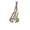
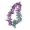
| Structure viewer | Molecule: MolmilJmol/JSmol |
|---|
- Downloads & links
-Download
| PDBx/mmCIF format | 7pgh.cif.gz | 231.1 KB | Display | PDBx/mmCIF format |
|---|---|---|---|---|
| PDB format | pdb7pgh.ent.gz | 189.5 KB | Display | PDB format |
| PDBx/mmJSON format | 7pgh.json.gz | Tree view | PDBx/mmJSON format | |
| Others |  Other downloads Other downloads |
-Validation report
| Arichive directory | https://data.pdbj.org/pub/pdb/validation_reports/pg/7pghftp://data.pdbj.org/pub/pdb/validation_reports/pg/7pgh | HTTPS FTP |
|---|
-Related structure data


-Links
 PDBj
PDBj


- Assembly
| Deposited unit | 
| ||||||||
|---|---|---|---|---|---|---|---|---|---|
| 1 |
| ||||||||
| Unit cell |
|
-Components
-Protein , 1 types, 8 molecules FABCDEGH
| #1: Protein | Mass: 16356.166 Da / Num. of mol.: 8 Source method: isolated from a genetically manipulated source Source: (gene. exp.) Alkalilimnicola ehrlichii (strain ATCC BAA-1101 / DSM 17681 / MLHE-1) (bacteria), (gene. exp.) Ruegeria pomeroyi (bacteria)Strain: ATCC BAA-1101 / DSM 17681 / MLHE-1 / Gene: Mlg_0322 / Production host: |
|---|
-Non-polymers , 5 types, 19 molecules 


| #2: Chemical | ChemComp-PE4 /  Mass: 354.436 Da / Num. of mol.: 9 / Source method: obtained synthetically / Formula: C16H34O8 / Comment: precipitant*YM Mass: 354.436 Da / Num. of mol.: 9 / Source method: obtained synthetically / Formula: C16H34O8 / Comment: precipitant*YM#3: Chemical | ChemComp-D12 /  Mass: 170.335 Da / Num. of mol.: 4 / Source method: obtained synthetically / Formula: C12H26 Mass: 170.335 Da / Num. of mol.: 4 / Source method: obtained synthetically / Formula: C12H26#4: Chemical | ChemComp-OCT / |  Mass: 114.229 Da / Num. of mol.: 1 / Source method: obtained synthetically / Formula: C8H18 Mass: 114.229 Da / Num. of mol.: 1 / Source method: obtained synthetically / Formula: C8H18#5: Chemical |  Mass: 546.646 Da / Num. of mol.: 3 / Source method: obtained synthetically / Formula: C24H50O13 / Comment: precipitant*YM Mass: 546.646 Da / Num. of mol.: 3 / Source method: obtained synthetically / Formula: C24H50O13 / Comment: precipitant*YM#6: Chemical |  Mass: 238.278 Da / Num. of mol.: 2 / Source method: obtained synthetically / Formula: C10H22O6 / Comment: precipitant*YM Mass: 238.278 Da / Num. of mol.: 2 / Source method: obtained synthetically / Formula: C10H22O6 / Comment: precipitant*YM |
|---|
-Details
| Has ligand of interest | N |
|---|---|
| Has protein modification | Y |
-Experimental details
-Experiment
| Experiment | Method: X-RAY DIFFRACTION / Number of used crystals: 1 |
|---|
- Sample preparation
| Crystal | Density Matthews: 4.94 Å3/Da / Density % sol: 75.11 % |
|---|---|
| Crystal grow | Temperature: 293 K / Method: vapor diffusion, hanging drop Details: Purified NaVAe1Sp1CTDp was concentrated to 13-13.5 mg ml-1 and crystallized in 22% PEG3350, 0.3 M KI, 8 mM sarcosine. Crystals were harvested in 30% PEG3350, 0.3 M KI, 8 mM sarcosine and 1mM ...Details: Purified NaVAe1Sp1CTDp was concentrated to 13-13.5 mg ml-1 and crystallized in 22% PEG3350, 0.3 M KI, 8 mM sarcosine. Crystals were harvested in 30% PEG3350, 0.3 M KI, 8 mM sarcosine and 1mM Fos-choline 12 (FC-12), Anatrace). |
-Data collection
| Diffraction | Mean temperature: 100 K / Serial crystal experiment: N | |||||||||||||||
|---|---|---|---|---|---|---|---|---|---|---|---|---|---|---|---|---|
| Diffraction source | Source: SYNCHROTRON / Site: APS / Beamline: 23-ID-B / Wavelength: 1, 0.97698, 0.97953, 0.95368 | |||||||||||||||
| Detector | Type: DECTRIS PILATUS 6M / Detector: PIXEL / Date: Apr 10, 2017 | |||||||||||||||
| Radiation | Protocol: MAD / Monochromatic (M) / Laue (L): M / Scattering type: x-ray | |||||||||||||||
| Radiation wavelength |
| |||||||||||||||
| Reflection | Resolution: 4.194→15 Å / Num. obs: 18983 / % possible obs: 96.53 % / Redundancy: 12.9 % / CC1/2: 0.999 / Rmerge(I) obs: 0.065 / Net I/σ(I): 11.6 | |||||||||||||||
| Reflection shell | Resolution: 4.194→4.342 Å / Rmerge(I) obs: 1 / Num. unique obs: 1766 / CC1/2: 0.404 |
- Processing
| Software |
| ||||||||||||||||||||||||||||||||||||||||||
|---|---|---|---|---|---|---|---|---|---|---|---|---|---|---|---|---|---|---|---|---|---|---|---|---|---|---|---|---|---|---|---|---|---|---|---|---|---|---|---|---|---|---|---|
| Refinement | Method to determine structure: MAD / Resolution: 4.194→14.989 Å / SU ML: 1.11 / Cross valid method: THROUGHOUT / σ(F): 1.34 / Phase error: 50.69 / Stereochemistry target values: ML
| ||||||||||||||||||||||||||||||||||||||||||
| Solvent computation | Shrinkage radii: 0.9 Å / VDW probe radii: 1.11 Å / Solvent model: FLAT BULK SOLVENT MODEL | ||||||||||||||||||||||||||||||||||||||||||
| Displacement parameters | Biso max: 650.8 Å2 / Biso mean: 317.5867 Å2 / Biso min: 30 Å2 | ||||||||||||||||||||||||||||||||||||||||||
| Refinement step | Cycle: final / Resolution: 4.194→14.989 Å
| ||||||||||||||||||||||||||||||||||||||||||
| LS refinement shell | Refine-ID: X-RAY DIFFRACTION / Rfactor Rfree error: 0
|
