Movie
Movie Controller

[English] 日本語
 Yorodumi
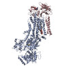
- PDB-7pem: Cryo-EM structure of phophorylated Drs2p-Cdc50p in a PS and ATP-b... -
+ Open data

- Basic information
| Entry | Database: PDB / ID: 7pem | |||||||||||||||||||||||||||
|---|---|---|---|---|---|---|---|---|---|---|---|---|---|---|---|---|---|---|---|---|---|---|---|---|---|---|---|---|
| Title | Cryo-EM structure of phophorylated Drs2p-Cdc50p in a PS and ATP-bound E2P state | |||||||||||||||||||||||||||
 Components Components |
| |||||||||||||||||||||||||||
 Keywords Keywords | MEMBRANE PROTEIN / Lipid Flippase / P4 ATPase / trans-Golgi Network / Phosphatidylserine transport | |||||||||||||||||||||||||||
| Function / homology |  Function and homology information Function and homology informationCdc50p-Drs2p complex / actin cortical patch localization / Ion transport by P-type ATPases / aminophospholipid translocation / phosphatidylcholine flippase activity / post-Golgi vesicle-mediated transport / phosphatidylserine flippase activity / phosphatidylserine floppase activity / ATPase-coupled intramembrane lipid carrier activity / phosphatidylethanolamine flippase activity ...Cdc50p-Drs2p complex / actin cortical patch localization / Ion transport by P-type ATPases / aminophospholipid translocation / phosphatidylcholine flippase activity / post-Golgi vesicle-mediated transport / phosphatidylserine flippase activity / phosphatidylserine floppase activity / ATPase-coupled intramembrane lipid carrier activity / phosphatidylethanolamine flippase activity / endocytic recycling / P-type phospholipid transporter / phosphatidylinositol-4-phosphate binding / retrograde transport, endosome to Golgi / phospholipid translocation / Neutrophil degranulation / trans-Golgi network / intracellular protein transport / endocytosis / late endosome membrane / endosome membrane / Golgi apparatus / magnesium ion binding / endoplasmic reticulum / ATP hydrolysis activity / ATP binding / plasma membrane / cytosol Similarity search - Function | |||||||||||||||||||||||||||
| Biological species |  | |||||||||||||||||||||||||||
| Method | ELECTRON MICROSCOPY / single particle reconstruction / cryo EM / Resolution: 3.1 Å | |||||||||||||||||||||||||||
 Authors Authors | Timcenko, M. / Wang, Y. / Lyons, J.A. / Nissen, P. / Lindorff-Larsen, K. | |||||||||||||||||||||||||||
| Funding support |  Denmark, 1items Denmark, 1items
| |||||||||||||||||||||||||||
 Citation Citation | Journal: To Be Published Title: Substrate Transport and Specificity in a Phospholipid Flippase Authors: Wang, Y. / Lyons, J.A. / Timcenko, M. / Kummerer, F. / de Groot, B.L. / Nissen, P. / Gapsys, V. / Lindorff-Larsen, K. | |||||||||||||||||||||||||||
| History |
|
- Structure visualization
| Structure viewer | Molecule: MolmilJmol/JSmol |
|---|
- Downloads & links
-Download
| PDBx/mmCIF format | 7pem.cif.gz | 289.3 KB | Display | PDBx/mmCIF format |
|---|---|---|---|---|
| PDB format | pdb7pem.ent.gz | 216.3 KB | Display | PDB format |
| PDBx/mmJSON format | 7pem.json.gz | Tree view | PDBx/mmJSON format | |
| Others |  Other downloads Other downloads |
-Validation report
| Arichive directory | https://data.pdbj.org/pub/pdb/validation_reports/pe/7pemftp://data.pdbj.org/pub/pdb/validation_reports/pe/7pem | HTTPS FTP |
|---|
-Related structure data
| Related structure data |  13353MC M: map data used to model this data C: citing same article ( |
|---|---|
| Similar structure data |
-Links
 PDBj
PDBj


- Assembly
| Deposited unit | 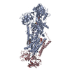
|
|---|---|
| 1 |
|
-Components
-Protein , 2 types, 2 molecules AC
| #1: Protein | Mass: 154006.766 Da / Num. of mol.: 1 Source method: isolated from a genetically manipulated source Details: Drs2p with a Thrombin cleavable C-terminal biotin acceptor domain (BAD) tag, and additional Thrombin cleavage site in the C-terminus. Source: (gene. exp.) Strain: ATCC 204508 / S288c / Gene: DRS2, YAL026C, FUN38 / Plasmid: pYeDP60 / Production host: References: UniProt: P39524, P-type phospholipid transporter |
|---|---|
| #2: Protein | Mass: 45037.312 Da / Num. of mol.: 1 Source method: isolated from a genetically manipulated source Details: Cdc50p with Thrombin cleavable C-terminal His-tag Source: (gene. exp.) Strain: ATCC 204508 / S288c / Gene: CDC50, YCR094W, YCR94W / Plasmid: pYeDP60 / Production host: |
-Sugars , 2 types, 3 molecules
| #3: Polysaccharide | Source method: isolated from a genetically manipulated source #4: Polysaccharide | beta-D-mannopyranose-(1-3)-beta-D-mannopyranose-(1-4)-2-acetamido-2-deoxy-beta-D-glucopyranose-(1-4) ...beta-D-mannopyranose-(1-3)-beta-D-mannopyranose-(1-4)-2-acetamido-2-deoxy-beta-D-glucopyranose-(1-4)-2-acetamido-2-deoxy-beta-D-glucopyranose | Source method: isolated from a genetically manipulated source |
|---|
-Non-polymers , 5 types, 6 molecules 


| #5: Chemical | ChemComp-2Y5 / ( Mass: 967.108 Da / Num. of mol.: 1 / Source method: obtained synthetically / Formula: C47H84O16P2 Mass: 967.108 Da / Num. of mol.: 1 / Source method: obtained synthetically / Formula: C47H84O16P2 |
|---|---|
| #6: Chemical | ChemComp-MG /  Mass: 24.305 Da / Num. of mol.: 1 / Source method: obtained synthetically / Formula: Mg Mass: 24.305 Da / Num. of mol.: 1 / Source method: obtained synthetically / Formula: Mg |
| #7: Chemical | ChemComp-ATP /  Mass: 507.181 Da / Num. of mol.: 1 / Source method: obtained synthetically / Formula: C10H16N5O13P3 / Comment: ATP, energy-carrying molecule*YM Mass: 507.181 Da / Num. of mol.: 1 / Source method: obtained synthetically / Formula: C10H16N5O13P3 / Comment: ATP, energy-carrying molecule*YM |
| #8: Chemical | ChemComp-Q3G /  Mass: 764.022 Da / Num. of mol.: 1 / Source method: obtained synthetically / Formula: C40H78NO10P / Feature type: SUBJECT OF INVESTIGATION / Comment: phospholipid*YM Mass: 764.022 Da / Num. of mol.: 1 / Source method: obtained synthetically / Formula: C40H78NO10P / Feature type: SUBJECT OF INVESTIGATION / Comment: phospholipid*YM |
| #9: Water | ChemComp-HOH / Mass: 18.015 Da / Num. of mol.: 2 / Source method: isolated from a natural source / Formula: H2O |
-Details
| Has ligand of interest | Y |
|---|---|
| Has protein modification | Y |
-Experimental details
-Experiment
| Experiment | Method: ELECTRON MICROSCOPY |
|---|---|
| EM experiment | Aggregation state: PARTICLE / 3D reconstruction method: single particle reconstruction |
- Sample preparation
| Component | Name: Binary complex of Drs2p-Cdc50p with the regulatory lipid PI4P and transport substrate PS Type: COMPLEX / Entity ID: #1-#2 / Source: RECOMBINANT | ||||||||||||||||||||||||||||||
|---|---|---|---|---|---|---|---|---|---|---|---|---|---|---|---|---|---|---|---|---|---|---|---|---|---|---|---|---|---|---|---|
| Molecular weight | Value: 0.18 MDa / Experimental value: NO | ||||||||||||||||||||||||||||||
| Source (natural) | Organism: | ||||||||||||||||||||||||||||||
| Source (recombinant) | Organism: | ||||||||||||||||||||||||||||||
| Buffer solution | pH: 7 | ||||||||||||||||||||||||||||||
| Buffer component |
| ||||||||||||||||||||||||||||||
| Specimen | Conc.: 0.6 mg/ml / Embedding applied: NO / Shadowing applied: NO / Staining applied: NO / Vitrification applied: YES Details: Purified in detergent lauryl maltose neopentyl glycol (LMNG) | ||||||||||||||||||||||||||||||
| Specimen support | Grid material: COPPER / Grid mesh size: 400 divisions/in. / Grid type: C-flat-1.2/1.3 | ||||||||||||||||||||||||||||||
| Vitrification | Instrument: FEI VITROBOT MARK IV / Cryogen name: ETHANE / Humidity: 100 % / Chamber temperature: 277.15 K Details: Incubated with 0.1 mg/mL POPS for 1 hour. 3mM ATP was added to the sample just before application to the grid. |
- Electron microscopy imaging
| Experimental equipment |  Model: Titan Krios / Image courtesy: FEI Company |
|---|---|
| Microscopy | Model: FEI TITAN KRIOS |
| Electron gun | Electron source:  FIELD EMISSION GUN / Accelerating voltage: 300 kV / Illumination mode: FLOOD BEAM FIELD EMISSION GUN / Accelerating voltage: 300 kV / Illumination mode: FLOOD BEAM |
| Electron lens | Mode: BRIGHT FIELD / Cs: 2.7 mm |
| Image recording | Average exposure time: 1.5 sec. / Electron dose: 60 e/Å2 / Film or detector model: GATAN K3 (6k x 4k) / Num. of grids imaged: 1 / Num. of real images: 9837 |
| EM imaging optics | Energyfilter name: GIF Bioquantum / Energyfilter slit width: 20 eV |
- Processing
| Software |
| |||||||||||||||||||||||||||||||||||||||||||||||||||||||
|---|---|---|---|---|---|---|---|---|---|---|---|---|---|---|---|---|---|---|---|---|---|---|---|---|---|---|---|---|---|---|---|---|---|---|---|---|---|---|---|---|---|---|---|---|---|---|---|---|---|---|---|---|---|---|---|---|
| EM software |
| |||||||||||||||||||||||||||||||||||||||||||||||||||||||
| CTF correction | Type: NONE | |||||||||||||||||||||||||||||||||||||||||||||||||||||||
| Particle selection | Num. of particles selected: 3029402 | |||||||||||||||||||||||||||||||||||||||||||||||||||||||
| Symmetry | Point symmetry: C1 (asymmetric) | |||||||||||||||||||||||||||||||||||||||||||||||||||||||
| 3D reconstruction | Resolution: 3.1 Å / Resolution method: FSC 0.143 CUT-OFF / Num. of particles: 91415 / Details: non-uniform refinement in cryoSPARC v3 / Symmetry type: POINT | |||||||||||||||||||||||||||||||||||||||||||||||||||||||
| Atomic model building | B value: 69.1 / Space: REAL / Target criteria: correlation coefficient Details: Molecular dynamics flexible fitting and energy minimization in Gromacs | |||||||||||||||||||||||||||||||||||||||||||||||||||||||
| Atomic model building | PDB-ID: 6ROJ Accession code: 6ROJ / Source name: PDB / Type: experimental model | |||||||||||||||||||||||||||||||||||||||||||||||||||||||
| Refinement | Cross valid method: NONE Stereochemistry target values: GeoStd + Monomer Library + CDL v1.2 | |||||||||||||||||||||||||||||||||||||||||||||||||||||||
| Displacement parameters | Biso mean: 61.24 Å2 | |||||||||||||||||||||||||||||||||||||||||||||||||||||||
| Refine LS restraints |
|