 ムービー
ムービー コントローラー


+ データを開く

- 基本情報
基本情報
| 登録情報 | データベース: PDB / ID: 7pee | |||||||||
|---|---|---|---|---|---|---|---|---|---|---|
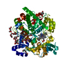
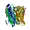
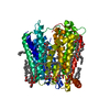
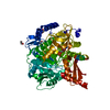
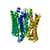
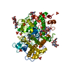
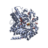
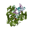
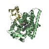
| タイトル | Crystal structure of extracellular part of human Trop2 | |||||||||
 要素 要素 | Tumor-associated calcium signal transducer 2 | |||||||||
 キーワード キーワード | MEMBRANE PROTEIN / extracellular part / dimer | |||||||||
| 機能・相同性 |  機能・相同性情報 機能・相同性情報negative regulation of branching involved in ureteric bud morphogenesis / ureteric bud morphogenesis / negative regulation of ruffle assembly / negative regulation of substrate adhesion-dependent cell spreading / negative regulation of epithelial cell migration / positive regulation of stem cell differentiation / regulation of epithelial cell proliferation / negative regulation of cell motility / negative regulation of stress fiber assembly / lateral plasma membrane ...negative regulation of branching involved in ureteric bud morphogenesis / ureteric bud morphogenesis / negative regulation of ruffle assembly / negative regulation of substrate adhesion-dependent cell spreading / negative regulation of epithelial cell migration / positive regulation of stem cell differentiation / regulation of epithelial cell proliferation / negative regulation of cell motility / negative regulation of stress fiber assembly / lateral plasma membrane / visual perception / basal plasma membrane / extracellular space / extracellular exosome / nucleus / membrane / cytosol 類似検索 - 分子機能 | |||||||||
| 生物種 |  Homo sapiens (ヒト) Homo sapiens (ヒト) | |||||||||
| 手法 |  X線回折 / シンクロトロン / 分子置換 / 解像度: 2.81 Å X線回折 / シンクロトロン / 分子置換 / 解像度: 2.81 Å | |||||||||
 データ登録者 データ登録者 | Pavsic, M. | |||||||||
| 資金援助 |  スロベニア, 2件 スロベニア, 2件
| |||||||||
 引用 引用 | ジャーナル: Int J Mol Sci / 年: 2021 タイトル: Trop2 Forms a Stable Dimer with Significant Structural Differences within the Membrane-Distal Region as Compared to EpCAM. 著者: Pavsic, M. | |||||||||
| 履歴 |
|
- 構造の表示
構造の表示
| 構造ビューア | 分子: MolmilJmol/JSmol |
|---|
- ダウンロードとリンク
-ダウンロード
| PDBx/mmCIF形式 | 7pee.cif.gz | 216.9 KB | 表示 | PDBx/mmCIF形式 |
|---|---|---|---|---|
| PDB形式 | pdb7pee.ent.gz | 159.3 KB | 表示 | PDB形式 |
| PDBx/mmJSON形式 | 7pee.json.gz | ツリー表示 | PDBx/mmJSON形式 | |
| その他 |  その他のダウンロード その他のダウンロード |
-検証レポート
| 文書・要旨 | 7pee_validation.pdf.gz | 1.2 MB | 表示 | wwPDB検証レポート |
|---|---|---|---|---|
| 文書・詳細版 | 7pee_full_validation.pdf.gz | 1.2 MB | 表示 | |
| XML形式データ | 7pee_validation.xml.gz | 34.5 KB | 表示 | |
| CIF形式データ | 7pee_validation.cif.gz | 47.1 KB | 表示 | |
| アーカイブディレクトリ | https://data.pdbj.org/pub/pdb/validation_reports/pe/7peeftp://data.pdbj.org/pub/pdb/validation_reports/pe/7pee | HTTPS FTP |
-関連構造データ
| 関連構造データ |  4mzvS S: 精密化の開始モデル |
|---|---|
| 類似構造データ |
-リンク
 PDBj
PDBj- 集合体
集合体
| 登録構造単位 | 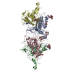
| ||||||||||||
|---|---|---|---|---|---|---|---|---|---|---|---|---|---|
| 1 | 
| ||||||||||||
| 2 | 
| ||||||||||||
| 単位格子 |
|
-要素
| #1: タンパク質 | 分子量: 28381.301 Da / 分子数: 4 / 変異: N120Q,N208Q / 由来タイプ: 組換発現 / 由来: (組換発現) Homo sapiens (ヒト) / 遺伝子: TACSTD2, GA733-1, M1S1, TROP2 / 細胞株 (発現宿主): Sf9発現宿主:   Spodoptera frugiperda (ツマジロクサヨトウ) Spodoptera frugiperda (ツマジロクサヨトウ)参照: UniProt: P09758 #2: 多糖 | #3: 化合物 |   分子量: 96.063 Da / 分子数: 2 / 由来タイプ: 天然 / 式: SO4 分子量: 96.063 Da / 分子数: 2 / 由来タイプ: 天然 / 式: SO4#4: 糖 |   タイプ: D-saccharide, beta linking / 分子量: 221.208 Da / 分子数: 2 / 由来タイプ: 天然 / 式: C8H15NO6 タイプ: D-saccharide, beta linking / 分子量: 221.208 Da / 分子数: 2 / 由来タイプ: 天然 / 式: C8H15NO6#5: 水 | ChemComp-HOH / |  分子量: 18.015 Da / 分子数: 30 / 由来タイプ: 天然 / 式: H2O 分子量: 18.015 Da / 分子数: 30 / 由来タイプ: 天然 / 式: H2O研究の焦点であるリガンドがあるか | N | Has protein modification | Y | |
|---|
-実験情報
-実験
| 実験 | 手法: X線回折 / 使用した結晶の数: 1 |
|---|
- 試料調製
試料調製
| 結晶 | マシュー密度: 5.05 Å3/Da / 溶媒含有率: 75.63 % / 解説: rod |
|---|---|
| 結晶化 | 温度: 294 K / 手法: 蒸気拡散法, シッティングドロップ法 / pH: 5.8 詳細: 1.6 M ammonium sulfate, 0.5 M NaCl, 0.0133 M EDTA, 0.1 M MES pH 5.8 |
-データ収集
| 回折 | 平均測定温度: 100 K / Serial crystal experiment: N |
|---|---|
| 放射光源 | 由来: シンクロトロン / サイト: ELETTRA  / ビームライン: 11.2C / 波長: 0.9789 Å / ビームライン: 11.2C / 波長: 0.9789 Å |
| 検出器 | タイプ: DECTRIS PILATUS 6M / 検出器: PIXEL / 日付: 2019年2月7日 |
| 放射 | プロトコル: SINGLE WAVELENGTH / 単色(M)・ラウエ(L): M / 散乱光タイプ: x-ray |
| 放射波長 | 波長: 0.9789 Å / 相対比: 1 |
| 反射 | 解像度: 2.81→48.38 Å / Num. obs: 57322 / % possible obs: 99.6 % / 冗長度: 6.6 % / Biso Wilson estimate: 64.21 Å2 / CC1/2: 0.998 / Rsym value: 0.1157 / Net I/σ(I): 15.14 |
| 反射 シェル | 解像度: 2.81→2.98 Å / Mean I/σ(I) obs: 1.85 / Num. unique obs: 9084 / CC1/2: 0.717 / Rsym value: 1.19 / % possible all: 99.4 |
- 解析
解析
| ソフトウェア |
| |||||||||||||||||||||||||||||||||||||||||||||||||||||||||||||||||||||||||||||||||||||||||||||||||||||||||||||||||||||||||||||||||||||||||||||||||||
|---|---|---|---|---|---|---|---|---|---|---|---|---|---|---|---|---|---|---|---|---|---|---|---|---|---|---|---|---|---|---|---|---|---|---|---|---|---|---|---|---|---|---|---|---|---|---|---|---|---|---|---|---|---|---|---|---|---|---|---|---|---|---|---|---|---|---|---|---|---|---|---|---|---|---|---|---|---|---|---|---|---|---|---|---|---|---|---|---|---|---|---|---|---|---|---|---|---|---|---|---|---|---|---|---|---|---|---|---|---|---|---|---|---|---|---|---|---|---|---|---|---|---|---|---|---|---|---|---|---|---|---|---|---|---|---|---|---|---|---|---|---|---|---|---|---|---|---|---|
| 精密化 | 構造決定の手法: 分子置換 開始モデル: 4MZV 解像度: 2.81→48.37 Å / SU ML: 0.4202 / 交差検証法: FREE R-VALUE / σ(F): 1.34 / 位相誤差: 28.8396 立体化学のターゲット値: GeoStd + Monomer Library + CDL v1.2
| |||||||||||||||||||||||||||||||||||||||||||||||||||||||||||||||||||||||||||||||||||||||||||||||||||||||||||||||||||||||||||||||||||||||||||||||||||
| 溶媒の処理 | 減衰半径: 0.9 Å / VDWプローブ半径: 1.11 Å / 溶媒モデル: FLAT BULK SOLVENT MODEL | |||||||||||||||||||||||||||||||||||||||||||||||||||||||||||||||||||||||||||||||||||||||||||||||||||||||||||||||||||||||||||||||||||||||||||||||||||
| 原子変位パラメータ | Biso mean: 75.8 Å2 | |||||||||||||||||||||||||||||||||||||||||||||||||||||||||||||||||||||||||||||||||||||||||||||||||||||||||||||||||||||||||||||||||||||||||||||||||||
| 精密化ステップ | サイクル: LAST / 解像度: 2.81→48.37 Å
| |||||||||||||||||||||||||||||||||||||||||||||||||||||||||||||||||||||||||||||||||||||||||||||||||||||||||||||||||||||||||||||||||||||||||||||||||||
| 拘束条件 |
| |||||||||||||||||||||||||||||||||||||||||||||||||||||||||||||||||||||||||||||||||||||||||||||||||||||||||||||||||||||||||||||||||||||||||||||||||||
| LS精密化 シェル |
|
