- PDB-3n9y: Crystal structure of human CYP11A1 in complex with cholesterol -
+
Open data
ID or keywords:
Loading...
-
Basic information
Entry
Database: PDB / ID: 3n9y
Title
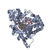
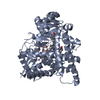
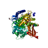
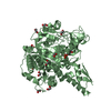
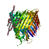
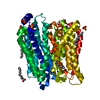
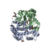
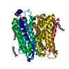
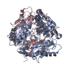
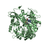
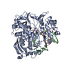
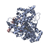
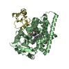
Crystal structure of human CYP11A1 in complex with cholesterol
Components
Adrenodoxin
Cholesterol side-chain cleavage enzyme
Keywords
Oxidoreductase / Electron transport / cytochrome P450 / cholesterol side chain cleavage / Structural Genomics / Structural Genomics Consortium / SGC
Function / homology
Function and homology information
steroid hormone biosynthetic process / cholesterol monooxygenase (side-chain-cleaving) / cholesterol monooxygenase (side-chain-cleaving) activity / cortisol metabolic process / Electron transport from NADPH to Ferredoxin / Defective CYP11A1 causes AICSR / glucocorticoid biosynthetic process / Mitochondrial iron-sulfur cluster biogenesis / hormone biosynthetic process / P450-containing electron transport chain ...steroid hormone biosynthetic process / cholesterol monooxygenase (side-chain-cleaving) / cholesterol monooxygenase (side-chain-cleaving) activity / cortisol metabolic process / Electron transport from NADPH to Ferredoxin / Defective CYP11A1 causes AICSR / glucocorticoid biosynthetic process / Mitochondrial iron-sulfur cluster biogenesis / hormone biosynthetic process / P450-containing electron transport chain / Protein lipoylation / sterol metabolic process / steroid biosynthetic process / C21-steroid hormone biosynthetic process / Pregnenolone biosynthesis / vitamin D metabolic process / cellular response to peptide hormone stimulus / Endogenous sterols / cholesterol metabolic process / cellular response to cAMP / cellular response to forskolin / electron transport chain / 2 iron, 2 sulfur cluster binding / electron transfer activity / mitochondrial inner membrane / iron ion binding / mitochondrial matrix / heme binding / mitochondrion Similarity search - Function
In the structure databanks used in Yorodumi, some data are registered as the other names, "COVID-19 virus" and "2019-nCoV". Here are the details of the virus and the list of structure data.
Jan 31, 2019. EMDB accession codes are about to change! (news from PDBe EMDB page)
EMDB accession codes are about to change! (news from PDBe EMDB page)
The allocation of 4 digits for EMDB accession codes will soon come to an end. Whilst these codes will remain in use, new EMDB accession codes will include an additional digit and will expand incrementally as the available range of codes is exhausted. The current 4-digit format prefixed with “EMD-” (i.e. EMD-XXXX) will advance to a 5-digit format (i.e. EMD-XXXXX), and so on. It is currently estimated that the 4-digit codes will be depleted around Spring 2019, at which point the 5-digit format will come into force.
The EM Navigator/Yorodumi systems omit the EMD- prefix.
Related info.:Q: What is EMD? / ID/Accession-code notation in Yorodumi/EM Navigator
Yorodumi is a browser for structure data from EMDB, PDB, SASBDB, etc.
This page is also the successor to EM Navigator detail page, and also detail information page/front-end page for Omokage search.
The word "yorodu" (or yorozu) is an old Japanese word meaning "ten thousand". "mi" (miru) is to see.
Related info.:EMDB / PDB / SASBDB / Comparison of 3 databanks / Yorodumi Search / Aug 31, 2016. New EM Navigator & Yorodumi / Yorodumi Papers / Jmol/JSmol / Function and homology information / Changes in new EM Navigator and Yorodumi
 Movie
Movie Controller


 Open data
 Basic information
 Components
 Keywords
 Function and homology information
 Homo sapiens (human)
 X-RAY DIFFRACTION /
  Authors
 Citation
 Structure visualization
 Downloads & links
 Other downloads

 PDBj
PDBj


 Assembly


 Mass: 616.487 Da / Num. of mol.: 2 / Source method: obtained synthetically / Formula: C34H32FeN4O4
 Mass: 386.654 Da / Num. of mol.: 2 / Source method: obtained synthetically / Formula: C27H46O
 Mass: 175.820 Da / Num. of mol.: 2 / Source method: obtained synthetically / Formula: Fe2S2
 Sample preparation
 / Beamline: 19-ID
 Processing